
Abstract
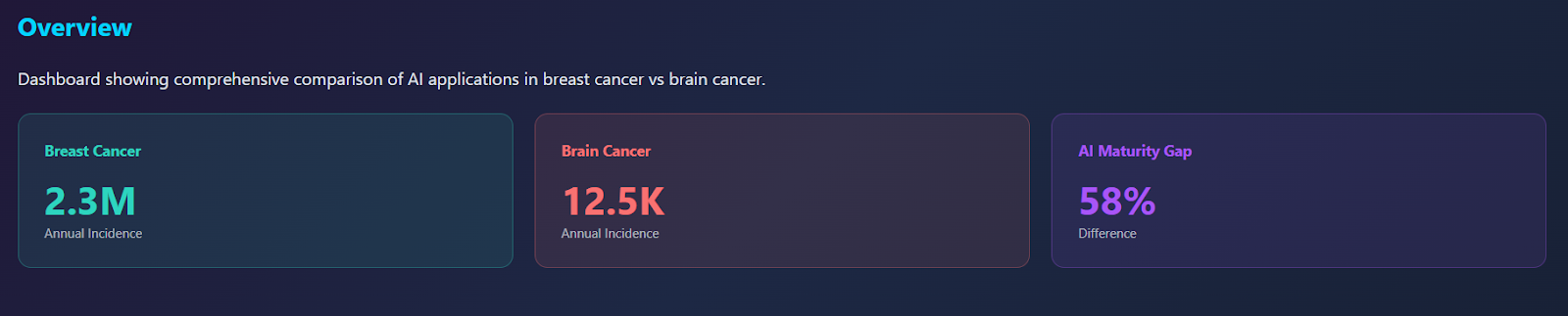
Breast cancer and brain cancer represent distinct malignancies with different epidemiologies – breast cancer affects over 2.3 million people annually worldwide, while brain cancer, though rarer, carries significantly higher mortality rates – molecular characteristics and treatment responses. Artificial intelligence (AI) has emerged as a promising clinical tool in oncology, though its maturity and applicability differ substantially between these two diseases. This review explores the differences and similarities in AI applications for breast cancer and brain cancer diagnosis and treatment. We examine how AI improves diagnostic accuracy, treatment planning and clinical decision making in both malignancies. Key findings indicate that AI demonstrates high clinical maturity in breast cancer applications due to standardised imaging protocols and large screening datasets, whereas brain cancer AI remains predominantly research-focused due to tumour complexity and variable MRI acquisition protocols. While both cancers benefit from enhanced image analysis and improved workflow efficiency, AI applications diverge in their specific uses: breast cancer prioritises AI-assisted screening and pathology analysis, whereas brain cancer focuses on surgical planning, tumour segmentation and radiogenomic prediction. This distinction reflects the unique biological and clinical challenges inherent to each malignancy. The appropriate extent of AI integration should therefore be disease-specific, with broader clinical implementation recommended for breast cancer diagnostics and more cautious, research-guided approaches for brain cancer. Successful integration requires robust validation, adaptive regulatory frameworks and sustained multidisciplinary collaboration between clinicians, data scientists and policymakers. These findings have direct implications for guiding future investment, clinical trial design and policy development in AI-driven oncology care.
Keywords: artificial intelligence, breast cancer, brain cancer, diagnosis, treatment, machine learning, clinical decision support.
Introduction
Cancer represents one of the most complex and diverse groups of diseases in modern medicine, characterised by the loss of normal regulatory control over cell growth and division. Under healthy conditions, cells proliferate only in response to specific signals and have mechanisms such as cell cycle checkpoints, DNA repair and apoptosis to ensure that damaged or unnecessary cells are eliminated (Alberts et al., 2022). Despite significant advancements in early detection and treatment, cancer remains a major global health challenge, accounting for nearly ten million deaths annually (World Health Organisation, 2025). Among the wide spectrum of cancer types, breast cancer and brain cancer stand out not only for their clinical importance but also for their distinct challenges.
Breast cancer is the most commonly diagnosed cancer in women worldwide (Sung et al., 2021). It typically originates in the epithelial cells of the ducts or lobules of the breast where a combination of hormonal, genetic and environmental factors can drive malignant transformation. On a more molecular level, breast cancer development is usually tied closely to dysregulation of systems controlled by the hormones oestrogen and progesterone, which normally regulate cell multiplication and differentiation in breast tissue (Thakur et al., 2022). Genetic mutations such as BRCA1, BRCA2, PIK3CA and TP53 can disrupt DNA repair and cell-cycle control, increasing susceptibility to breast cancer (Wang et al., 2024). Breast cancers are commonly classified by the presence of ER, PR and HER2 receptors (Beecher et al., 2022). These subtypes guide treatment: ER-positive tumours respond to endocrine therapy, HER2-positive tumours to targeted agents like trastuzumab, while triple-negative cancers, lacking all three receptors, are typically more aggressive and harder to treat (Ross & Fletcher, 2023; Lehmann et al., 2022). Clinically, breast cancer is frequently detected through imaging, particularly mammography, which has been used in screening programmes for several decades, due to the structural organisation of breast tissue and the high prevalence of the disease. As a result, massive datasets containing millions of labelled mammograms, coupled with detailed pathology reports and long term clinical outcomes have been accumulated over time (Breast Cancer Research 2023). This combination of biological characterisation and imaging standardisation has positioned breast cancer as one of the most data rich and analytically mature areas of medical research (McKinney et al., 2020). Consequently, it provides the ideal environment for training, validating and integrating artificial intelligence (AI) systems into routine clinical care, with algorithms benefitting from the scale, consistency and quality of available data.
Brain cancer represents a smaller but far more biologically intricate group of malignancies, arising either as primary tumours, most commonly gliomas, meningiomas and medulloblastomas, or as secondary brain metastases from cancers elsewhere. Primary malignant brain tumours, particularly glioblastoma, originate from glial cells and are characterised by extreme genetic instability, rapid multiplication and highly invasive growth that takes over healthy brain tissue (Neuro-Oncology Insights, 2023). At the molecular level, brain tumours are shaped by mutations affecting cell differentiation, metabolism and DNA repair such as IDH1/2 mutations, MGMT promoter methylation, EGFR amplification and PTEN loss, which influence prognosis and treatment response (Gupta et al., 2024). For instance, IDH-mutant gliomas are slower-growing and more treatment-sensitive, while IDH-wild-type glioblastomas are typically more aggressive and resistant to therapy (Weller et al., 2021). Unlike breast cancer, brain tumours lack widely implemented population screening programmes, partly due to their lower incidence and the sensitive anatomical environment in which they arise (Louis et al., 2021). As a result, many cases present only once symptoms develop, often at a later stage. Data collection is therefore more limited, and biological diversity across tumour types, combined with a smaller dataset makes brain cancer a more challenging area for large scale research. The complexity and unmet clinical need provide strong motivation for advanced AI systems that can support diagnosis, characterisation and treatment planning.
Breast and brain cancers share many challenges such as treatment resistance, tumour heterogeneity and risk of recurrence, but the nature of these challenges differs a lot. Breast cancer is often more treatable due to earlier detection and more targeted therapies, whereas brain cancer remains far more difficult because of its location, biological complexity and barriers like the blood-brain barrier. However, artificial intelligence offers a unifying technological approach that can support diagnosis and treatment across both areas. AI has emerged as a transformative force in healthcare, particularly within diagnostics, imaging and clinical decision support (Ogut, 2025). Encompassing machine learning (ML), deep learning (DL), natural language processing (NLP) and predictive analytics, AI’s value in oncology lies in its ability to analyse enormous quantities of medical data, such as imaging, genomic information and electronic health records, and detect patterns that clinicians may not readily see (Esteva et al., 2021). In recent years, AI has demonstrated promising capabilities in early cancer detection, tumour segmentation, treatment optimisation through personalised planning and even the prediction of patient outcomes. Rather than replacing clinicians, these systems aim to improve clinical judgement, reduce diagnostic variability and improve overall efficiency. Within breast and brain cancer, AI has been applied in distinct but equally important ways. In breast cancer, deep learning algorithms have shown performance comparable to, and sometimes exceeding, that of radiologists in identifying early lesions on mammograms (Yale et al., 2022). AI models can classify breast density, highlight suspicious regions and reduce both false-positive and false-negative rates, thereby supporting radiologists in screening programmes. Predictive models also help personalise treatment by estimating recurrence risk, assessing chemotherapy benefit and informing long-term planning (Oncology AI Review, 2023). In brain cancer, where imaging is more complex, AI applications are more experimental. One rapidly advancing area is automated tumour segmentation on MRI, a task traditionally done manually by neuroradiologists. Accurate segmentation is essential for surgical planning, radiotherapy targeting and monitoring tumour progression or treatment response. AI-driven operative tools, such as navigation systems and real-time tissue classification, are also emerging to help neurosurgeons maximise tumour resection while preserving neurological function (Swinburne et al., 2022).
Despite AI’s rapidly expanding potential, its integration into healthcare raises important questions regarding ethics, and more specifically, bias, reproducibility and accountability. These concerns become particularly relevant when comparing two cancer types with very different datasets and clinical structures. Breast cancer’s long established screening systems provide a consistent and wide source of data, making it easier to train and evaluate AI models. Conversely, brain cancer’s complexity, rareness and imaging variability pose significant challenges to achieving the same level of clinical integration. As a result, while AI in breast cancer is progressing into routine clinical practice, many brain cancer AI tools remain confined to research settings or early stage trials. These distinctions make breast and brain cancer ideal contrasting case studies for evaluating the broader question guiding this research paper; by examining their similarities and differences, we can better understand both the opportunities and limitations of AI across oncology as a whole.
Diagnosis and AI in Brain Cancer
Diagnosis of brain cancer relies heavily on MRI due to the complex structure of the brain and the infiltrative nature of these tumours. In particular, this is true for gliomas and glioblastomas which are cancers that arise from glial cells – the supportive cells of the brain. Gliomas range in severity, while glioblastomas represent the most aggressive form, making accurate imaging especially important (Weller et al., 2021).
Traditional radiological assessment depends on two-dimensional measurements and subjective interpretation; however, this approach can lead to considerable differences between observers. As a result, AI has emerged as a powerful tool in this context, mainly through automated tumour segmentation and radiomics-based analysis. For example, deep learning models can outline the enhancing tumour, non-enhancing core and surrounding oedema with accuracy comparable to expert neuroradiologists (Isensee et al., 2021). This is important because automated segmentation provides consistent volumetric measurements, enabling clinicians to detect subtle changes in tumour size that may not be easily visible and therefore monitor treatment response more reliably.
AI has also demonstrated promise in the non-invasive prediction of molecular tumour characteristics, an increasingly important component of brain tumour diagnosis following the 2021 WHO CNS classification update (Louis et al., 2021). Radiogenomic AI models have been shown to infer key markers such as IDH mutation status, MGMT promoter methylation and 1p/19q co-deletion from MRI scans (Weller et al., 2021). These markers strongly influence prognosis and treatment selection; for example, MGMT-methylated glioblastomas respond better to alkylating chemotherapy, while IDH-mutant gliomas have distinctly different survival trajectories. AI-driven prediction therefore provides an avenue for faster, less invasive diagnostic workflows, reducing reliance on surgical biopsies where imaging may already contain this molecular information.
Beyond classification, AI enhances diagnostic precision by reducing intra- and inter-observer variability. An international multi-reader study showed that AI-assisted tumour response assessment significantly improved reproducibility among clinicians evaluating MRI scans (Reina et al., 2022). Standardised assessments are particularly important for distinguishing true progression from pseudoprogression, a long-standing challenge in neuro-oncology caused by treatment-induced imaging changes. Additionally, natural language processing systems applied to radiology reports can detect subtle discrepancies or incomplete documentation, further reinforcing diagnostic quality (Liu et al., 2023).
Despite its promise, brain cancer AI faces considerable challenges. Variability in MRI acquisition protocols between hospitals reduces model generalisability. Moreover, the rarity of certain tumour subtypes limits the availability of large, annotated datasets (Neuro-Oncology Insights, 2023). As a result, many AI tools for brain tumour diagnosis remain in the research phase or early validation trials rather than being deployed clinically on a large scale.
Treatment and AI in Brain Cancer
AI is increasingly contributing to treatment planning and prognostication in neuro-oncology. For neurosurgical intervention, deep learning models assist in defining optimal resection margins while preserving eloquent brain areas. This is essential because more extensive resection correlates with improved survival in glioblastoma, but aggressive surgery risks damage to critical structures. AI-based navigation systems and intraoperative tissue classification tools can therefore support neurosurgeons in differentiating tumours from healthy tissue in real time (Swinburne et al., 2022).
AI also plays a growing role in radiotherapy planning. Automated segmentation accelerates preparation for radiotherapy by producing consistent contouring of tumour and oedema volumes, tasks that are time-consuming when performed manually. Standardised contours reduce planning differences between clinicians, which can significantly affect dose distribution and treatment efficacy.
In systemic therapy, machine learning algorithms analysing multimodal data, including MRI, genetics and clinical variables, can predict survival trajectories and treatment responses, helping clinicians identify the most appropriate therapeutic pathway for individual patients (Weller et al., 2021). Radiomic signatures, for example, have been associated with chemotherapy responsiveness and expected disease progression rates. These data-driven predictions may eventually support personalised therapy planning, although most models require further external validation.
Nonetheless, clinical deployment of AI in brain cancer treatment remains limited by regulatory, ethical and data-quality concerns. The lack of large multicentre trials evaluating AI-driven treatment planning restricts translation into routine care. In addition, clinicians emphasise the need for explainable models to ensure safety and trust (Reina et al., 2022).
Diagnosis and AI in Breast Cancer
Breast cancer diagnosis benefits from decades of structured screening programmes and highly standardised imaging, particularly mammography. These conditions have produced vast annotated datasets, enabling rapid development and validation of AI systems. Deep learning algorithms have demonstrated diagnostic performance comparable to, and sometimes exceeding, radiologists in detecting mammographic abnormalities (McKinney et al., 2020). Multiple studies show that AI can reduce false positives and false negatives, highlight suspicious regions and classify breast density, an important risk factor for breast cancer that affects imaging sensitivity (Yala et al., 2022).
More recent systematic reviews show that AI-assisted readings often match or exceed the performance of the traditional double-reading workflow used in screening settings, while reducing radiologist workload without compromising diagnostic quality (Zhao et al., 2023). AI systems can act as a second reader, a decision-support tool or a triage mechanism that prioritises abnormal scans for human review. These flexible modes of integration allow AI to enhance efficiency while maintaining clinical safety.
AI is also having a growing impact on risk prediction. Image-only deep learning models trained on mammograms have demonstrated significantly higher accuracy for predicting future breast cancer risk than traditional risk calculators such as the Gail or Tyrer-Cuzick models (Yala et al., 2022). By leveraging subtle patterns not visible to the human eye, AI can identify women who may benefit from more frequent screening or supplemental imaging such as MRI. Studies have reported area-under-the-curve (AUC) values ranging from 0.72 to 0.90 for these models, outperforming conventional risk tools that typically achieve AUCs around 0.60 (Zhao et al., 2023).
Furthermore, AI-enhanced ultrasound and MRI applications improve the detection of lesions in dense breast tissue, a known limitation of mammography. By increasing the visibility of subtle abnormalities, these tools help radiologists interpret images more quickly and with greater confidence, thereby improving overall diagnostic efficiency. Additionally, radiomics models can differentiate benign from malignant lesions more accurately, potentially reducing unnecessary biopsies (Breast Cancer Research, 2023). Similarly to brain tumours, NLP tools applied to breast imaging reports support standardisation and reduce reporting inconsistencies (Liu et al., 2023).
Treatment and AI in Breast Cancer
AI supports multiple aspects of breast cancer treatment planning and management. Predictive models analysing imaging, pathology and genomic data can estimate chemotherapy benefit, recurrence likelihood and response to neoadjuvant therapy (Oncology AI Review, 2023). For example, AI models that combine radiomics with gene-expression profiles have shown strong accuracy for predicting pathological complete response to chemotherapy, enabling more personalised treatment approaches.
AI also plays a role in guiding surgical management. Deep learning algorithms can assist in preoperative planning by assessing tumour extent more accurately on MRI, supporting decisions such as breast-conserving surgery versus mastectomy (Ross & Fletcher, 2023). In radiation oncology, AI streamlines contouring of tumour beds and organs at risk, improving consistency across clinicians and reducing preparation time.
Pathology workflows benefit significantly from AI-based image analysis. Digital pathology algorithms can identify mitotic figures, grade tumours and quantify hormone receptor expression with high accuracy, reinforcing pathologist decision making (Genomic Predictive Biomarkers, 2022). This enhances reproducibility and reduces diagnostic delays.
As with brain cancer, ethical and regulatory challenges remain. Ensuring equitable performance across demographic groups, securing patient data and guaranteeing transparency of model decisions are essential for responsible use. Nevertheless, breast cancer AI, supported by large datasets and consistent imaging, has already reached a higher clinical maturity level than brain cancer applications.
Analysis: Effectiveness of AI in Brain and Breast Cancer
The integration of AI into cancer care reflects the distinct structural, biological and data-related differences between brain and breast cancer. Breast cancer benefits immensely from large-scale screening programmes and standardised mammographic imaging, providing fertile ground for training and validating robust AI systems. As a result, AI use in breast cancer screening and diagnostics is rapidly transitioning into routine practice. In contrast, brain cancer presents greater biological heterogeneity, imaging protocols that vary between hospitals and scanners, and limited dataset sizes, all of which slow AI’s transition into clinical settings. Nonetheless, AI’s strengths in neuro-oncology, especially segmentation, surgical planning and radiogenomic inference, address the unique and challenging aspects of brain tumour management.
As cancerous tumours in the brain and breasts evolve and mutate, they become more complex and are at higher risk of metastasing. It is pivotal to detect them earlier on and while they are still an early stage cancer (Stages 1 and 2), which is commonly staged by a number staging system or the TNM (Tumour, Node, Metastasis) system (Cancer Research UK, 2023). The emergence of artificial intelligence-based tools in medical imaging has been motivated by the desire for greater efficiency and efficacy in clinical care.
By analysing vast numbers of mammograms, ultrasounds and MRI images, artificial intelligence systems learn what normal and cancer-indicating scans look like, helping radiologists spot subtle signs of disease that might otherwise go unnoticed. AI may also reduce unnecessary biopsies by lowering false positives and flag individuals at higher risk earlier. In a large randomised controlled trial, 80,033 women aged between 40 and 80 years old were assigned either a standard reading of screening mammograms or an AI-supported protocol using the Transpara system (Lång et al., 2023). Artificial intelligence, utilised by doctors and radiologists, safely demonstrated during the clinical breast cancer trial that AI-assisted mammogram readings, compared to standard double readings, detect more cancers (6.1 vs 5.1 per 1000), do not increase false negatives and reduce the radiologist screen-reading workload by approximately 44%. The authors from this trial also concluded that AI-supported screening is clinically safe (Lång et al, 2023). There are multiple public mammography datasets (DDSM/CBIS-DDSM, MIAS, INBreast, OPTIMAM and others) – plus routine screening programmes – that have enabled the large-scale training and development of AI within breast cancer diagnosis (El Karawi et al., 2024). Due to the substantial number of accessible mammograms and screening data, AI can be implemented effectively in the detection and location of cancerous tumours in the breasts.
It is strongly evident that AI has reached clinical maturity in breast cancer imaging; on the other hand, this contrasts with AI being utilised in brain cancer detection, which currently seems to be less effective.
Malignant brain tumours can be detected by CT and MRI scans, in which AI may be leveraged for protocol development, imaging acquisition, reconstruction, interpretation and clinical care (Paudyal et al., 2023). However, cancerous brain tumours are far more complex and less common than breast cancer. In the UK, there are approximately 13,000 new cases of brain, other CNS and intracranial tumours each year, which accounts for only 3% of all new cancer cases (Cancer Research UK, 2025). Since it is not common, there is inadequate research and fewer standardised, large datasets available for testing and validating the incorporation of AI in CT and MRI scans. This acts as a limiting factor for the active development of AI-assisted tools in brain cancer and restricts its application by keeping it at the research stage rather than in clinical use. Additionally, the high tumour heterogeneity observed in brain cancers is another significant obstacle. Brain tumours exhibit significant variation in size, shape, internal structure and imaging appearance even within a single tumour type, and they comprise numerous distinct histological types (gliomas, meningiomas, metastases, lymphomas, etc.). For AI models trained on small or homogeneous datasets, these biological differences result in variable signal intensities and ambiguous borders on MRI (and frequently limited soft-tissue contrast on CT), making automated detection, classification and segmentation much more difficult (Cè et al., 2023).
Practical issues, such as missing MRI sequences, variable slice thicknesses and inconsistent pre-processing, may further reduce the development of AI in malignant brain tumour detection as it is too inconsistent for the AI-supported scanner to create classification methods, trends and patterns like in breast cancer mammography. The use of AI-supported scanning in brain cancer is disrupted by the wide variations in MRI scanner type in each hospital location, field strength, pulse sequences and imaging protocols between hospitals. An AI-assisting model trained on data from one medical institution may not assist effectively when applied to data from another due to the inter-site differences, which causes a “domain shift” (Dorfner et al., 2025). Important MRI sequences may also be absent or inconsistent among patients in real-world clinical settings. For instance, a segmentation study discovered that models trained with “sequence dropout” (eg., reliability to missing MRI modalities) performed noticeably better on incomplete, large-scale data (Ruffle et al., 2023). While AI shows promise through its precision and accuracy in research settings, most work in brain cancer remains research-focused until data trends and clinical workflows are solved.
Therefore, AI has been a great help in the early detection of both breast and brain cancers, but its effectiveness is very different in these two contexts. For instance, breast cancer mammography has public access to well-established imaging techniques, large and consistent screening datasets, and successful clinical trials which together enable AI-supported screenings to perform reliably in everyday practice. On the other hand, AI for brain cancer is still mainly focused on research due to the infrequency and biological complexity of brain tumours, along with marked differences in MRI acquisition among hospitals. These factors reduce the generalisation of the model, which makes clinical adoption slower and less efficient in neuro-oncology than in breast imaging.
Surgical and Machinery Approach to Tackling Brain and Breast Cancer
Artificial intelligence is advancing in the field of oncology. While the overall goal is to enhance image analysis, streamline workflows and boost diagnostic accuracy, which applies to various cancer types, the specific clinical focus for AI can vary quite a bit from one cancer to another. For example, in breast cancer, AI is predominantly aimed at screening methods such as mammography and pathology evaluation; on the other hand, in cases like brain cancer, particularly gliomas, the key contributions of AI are found in areas like surgical planning, tumour segmentation and radio genomic predictions. The following section will dive into these distinct applications, shedding light on how they stem from the unique biological and clinical traits of each type of cancer, and explore what this means for patient care.
Across different cancer types, AI presents several common benefits. For example, AI models – especially those powered by deep learning – can quickly process imaging data, uncover subtle features that might escape the human eye and help minimise variability between different observers. In regards to breast cancer screening, AI can aid radiologists by highlighting suspicious areas on mammograms, potentially lightening their workload and enhancing cancer detection rates (McKinney et al., 2020). Likewise, in brain cancer, AI-assisted segmentation of MRI scans can lead to more accurate tumour boundary definitions, thereby improving consistency in surgical planning and radiotherapy treatment (Tustison et al., 2020).
Despite these shared advantages, the most impactful applications of AI reveal distinct challenges within the diagnostic and treatment pathways. These variations originate from the fact that breast cancer is frequently identified and treated through population screening and pathology, whereas brain tumours demand more intricate neurosurgical and molecular tactics, which require a delicate touch and careful handling.
One of the earliest and most widely studied applications of AI in breast cancer is mammography-based screening. A review of test-accuracy studies found that most AI systems, when evaluated on large screening datasets, were less accurate than a single radiologist and notably worse than consensus readings by two radiologists (Freeman et al., 2021). The authors concluded that, although AI holds promise, current systems are not yet specific enough to replace radiologist double-reading in population-level screening. Another emerging direction is the reduction of risk through AI prediction models. Rather than just detecting cancer, these models estimate a woman’s likelihood of developing cancer in future years, guiding tailored follow up and potentially reducing unnecessary imaging (Strand, 2025). For example, risk models based on mammographic density which rely on the help of deep-learning-derived features have been proposed to inform recall decisions or direct women to supplemental imaging like MRI or ultrasound based on elevated risk.
In pathology, AI is revolutionising how breast tissue specimens are analysed. With the digitisation of glass slides, convolutional neural networks (CNNs) and other techniques have been applied to tasks such as hormone receptor quantification (ER, PR), HER2 detection, Ki‑67 indexing and mitotic figure counting (Chan et al., 2023). These tools not only speed up the pathology workflow but also increase objectivity and reproducibility across laboratories. A literature review highlighted that AI can help assess tumour-infiltrating lymphocytes (TILs), identify lymph node metastases and more reliably report biomarker expression – tasks that are otherwise time-consuming and subject to the variability of two people. In particular, a 2024 review emphasised how AI-powered quantitative biomarker analysis (e.g., ER/PR/HER2) improves inter-observer concordance, thereby supporting more consistent decision making (Soliman et al., 2024).
Moreover, AI in pathology is not limited to diagnosis: predictive models trained on histological data can forecast treatment response (eg., to neoadjuvant chemotherapy) and prognosis, enabling more personalised care according to (Uchikov et al., 2024)
Brain tumours, particularly gliomas, pose unique challenges. They infiltrate healthy tissue, vary in grade and molecular subtype, and reside in a highly constrained anatomical context. AI has been applied to preoperative MRI to segment the tumour, identify its spatial relationship to critical brain structures and assist with surgical planning (Irmici et al., 2023). In surgical settings, deep learning models can integrate imaging (eg., diffusion tensor imaging, functional MRI) to suggest optimal margins, balancing maximal tumour removal against functional preservation (Evangelou, 2025). A review on neurosurgery described how AI could augment intraoperative navigation, assist with margin assessment in augmented reality frameworks and even predict surgical outcomes (Williams et al., 2021). This integration supports a shift toward precision neurosurgery, where resection is tailored to both tumours and individual neuroanatomy.
Beyond segmentation, perhaps the most interesting area in brain cancer is radio genomics, linking imaging features with the tumour’s molecular and genetic makeup. AI-driven radiomics can extract large sets of quantitative features (texture, shape, intensity) from MRI, which can then be compared with genomic alterations (eg., IDH mutation status, 1p/19q codeletion) via AI models. These radio genomic models enable non-invasive molecular identification, reducing the reliance on tissue biopsy and potentially speeding up decision making. For example, in gliomas, AI-based MRI radio genomic models have been demonstrated to predict histological grade, survival and treatment response. Fan et al. (2024) provided a detailed workflow for such models, showing how feature extraction, selection and classification can translate to clinically actionable predictions (Fan et al., 2024). Another key application is tumour recurrence prediction. A systematic review of preoperative MRI-based AI models found that they can predict local and distant recurrence of gliomas, potentially guiding surgeons in deciding how wide a resection margin should be and where to escalate the radiotherapy dose (Bijlsma et al., 2025).
In addition to characterisation, AI models that integrate radiomic, clinical and molecular data offer powerful risk removal tools. These models can predict overall survival, reoccurrence risk and response to therapies such as temozolomide or radiotherapy (Khalighi et al., 2024). By simulating treatment outcomes, AI may enable truly individualised care, optimising dose and timing based on a patient’s predicted trajectory. Furthermore, for aggressive brain tumours like glioblastoma, rapid AI-driven molecular diagnosis may be possible using optical imaging combined with deep learning. For example, a recent study developed “Deep Glioma”, which uses stimulated Raman histology for rapid molecular subtype prediction (eg., IDH, 1p/19q) in under 90 seconds, achieving over 93% accuracy in a multicentre cohort (Hollon et al., 2023). This could dramatically shorten the time to take the diagnosis and facilitate care and therapy.
While AI’s applications are promising, there are significant challenges that must be addressed for clinical translation. These problems include the validation and stark differences between different analyses of AI. Many studies in both breast and brain cancer are different, from single and seperate centres, and use contrasting imaging protocols such as MRI, CT and ultrasound (Geppert et al.,2023). Models must be externally validated across institutions, scanners and patient populations. Another problem is the interpretability of the AI models. AI models, especially deep neural networks, often act as “black boxes” (a complex system where data is hidden and can only be understood by internal workings), raising concerns about trust, bias and accountability. In neuro-oncology, access to data is profoundly important and the restriction of certain data could result in false diagnoses, which would result in fatal and misleading treatments. Another issue is the ethical and data issues of AI. For both cancers, implementing AI in oncological care means going through data privacy regulations and the need for standardisation of imaging and pathology detection in all hospitals. Using different systems in different hospitals could cause issues in transferring and utilising data. In brain cancer, any sensitive information that is captured from the imaging must be deleted before radiologists have access to them. A solution could be to have an AI data analyser to ensure mammographical or imaging data is double-checked against an extensive database before radiologist analysis. The final issue that will be discussed is resource inequality. Access to AI may not be available in all countries perhaps due to lack of funds or the lack of sanitary conditions. Organisations such as the UN or WHO should start initiatives in developing countries to ensure world-class healthcare is accessible to all.
Conclusion
Artificial intelligence has demonstrated considerable potential to reshape cancer diagnosis and treatment, yet its impact varies significantly between breast and brain cancer because of fundamental differences in biology, data availability and clinical practice. Breast cancer, supported by decades of population-level screening, standardised mammography protocols and extensive publicly-available imaging datasets, has created an environment in which AI can be rigorously trained, externally validated and safely incorporated into routine workflows. As a result, AI tools in breast cancer, particularly those used for screening, risk prediction and pathology, have reached a level of maturity that allows for broad, clinically meaningful integration.
By contrast, AI development in brain cancer must contend with the inherent complexity of intracranial tumours, the lack of large harmonised datasets and substantial variability in MRI acquisition across institutions. These constraints limit the generalisability of current models and mean that most systems remain confined to research settings or early-stage clinical evaluation. Even so, the progress of AI in neuro-oncology, especially in tumour segmentation, surgical planning and radiogenomic characterisation, highlights its promise for improving some of the most technically challenging aspects of cancer care.
However, artificial intelligence is a brand new technological advancement, which means that there are still many implications in utilising it within the diagnosis and treatment of breast and brain cancers. A major risk of AI-assisted systems could be over-reliance, as doctors and radiologists may trust AI more than their own judgement. Another implication could be AI’s bias since it is trained and developed with previous data; as a result, it could lead to unequal care.
Taken together, these findings suggest that AI should be integrated into oncology in a disease-specific, evidence-driven manner. For breast cancer, widespread deployment is justified and likely to continue expanding as models improve and healthcare systems adapt. For brain cancer, however, cautious adoption is essential, prioritising multicentre validation, imaging standardisation and explainable model design before broad clinical implementation can be considered.
Looking ahead, strengthening data quality, improving interpretability and fostering collaboration between clinicians, engineers and regulatory bodies will be vital for realising AI’s full potential across both cancers. Ultimately, the comparison between breast and brain cancer illustrates a central truth in modern oncology: AI succeeds most rapidly when data is abundant and biology is more predictable, and progresses more gradually when disease is highly complex and patient populations are smaller, but in both settings it offers a path towards more precise and equitable care.
Bibliography
Abdel Razek, A.A.K., Alksas, A., Shehata, M., AbdelKhalek, A., Abdel Baky, K., El-Baz, A. & Helmy, E. (2021) ‘Clinical applications of artificial intelligence and radiomics in neuro-oncology imaging’, Insights into Imaging, 12, p.152.
Alberts, B., Johnson, A., Lewis, J., Raff, M., Roberts, K. & Walter, P. (2022) Molecular Biology of the Cell, 7th edn. New York: Garland Science.
Al-Karawi, D., Al-Zaidi, S., Helael, K.A. et al. (2024) ‘A review of artificial intelligence in breast imaging’, Tomography. 10(5), pp.705-726.
American Cancer Society (2024) Breast Cancer Facts & Figures 2024–2025 [online]. <https://www.cancer.org/content/dam/cancer-org/research/cancer-facts-and-statistics/breast-cancer-facts-and-figures/2024/breast-cancer-facts-and-figures-2024.pdf>
Beecher, K., Kulasegaran, T., Lakhani, S.R. & Reed, A.E.M. (2025) ‘Receptor-based classification of breast cancer’, International Journal of Molecular Sciences, 26(15).
Bijlsma, S., Maal, T., Rubbert, C. et al. (2025) AI radiomics predicts spatial glioma recurrence on preoperative MRI: a systematic review’, Eur J Radiol, 193:112412.
Breast Cancer Research (2023) ‘Mammography dataset development and clinical applications’, Breast Cancer Research, 25(4), pp.1–12.
Cancer Genomics Review (2024) ‘Molecular drivers of glioma: an updated overview’, Cancer Genomics Review, 18(2), pp.44–59.
Cancer Research UK (2023) Stages and grades of breast cancer, Cancer Research UK [online].<https://www.cancerresearchuk.org/about-cancer/breast-cancer/stages-grades>
Cancer Research UK (2023) What is Cancer?, Cancer Research UK [online]. <https://www.cancerresearchuk.org/about-cancer/what-is-cancer>
Cancer Research UK (n.d.) Brain, other CNS and intracranial tumours statistics (2025), Cancer Research UK [online]. <https://www.cancerresearchuk.org/health-professional/cancer-statistics/statistics-by-cancer-type/brain-other-cns-and-intracranial-tumours>
Cè, M., Menze, B.H., van Ginneken, B., Reyes, M. & Underwood, J. (2023) ‘Artificial intelligence in brain tumour imaging’, European Radiology Experimental, 7(1), 46.
Chan, R.C., To, C.K.T., Cheng, K.C.T. et al. (2022) ‘Artificial intelligence in breast cancer histopathology’, Histopathology, 82(1), pp.198-210.
Datwani, S., Khan, H., Niazi, M.K.K., Parwani, A.V. & Li, Z. (2025) ‘Artificial intelligence in breast pathology: overview and recent updates’, Human Pathology, 162:105819.
Dorfner, F.J., Patel, J.B., Kalpathy-Cramer, J., Gerstner, E.R. & Bridge, C.P. (2025) ‘A review of deep learning for brain tumour analysis in MRI’, npj Precision Oncology, 9(2).
Dur Karasayar, A.H., Kulaç, I. & Kapucuoğlu, N. (2025) ‘Advances in breast cancer care: the role of artificial intelligence and digital pathology in precision medicine’, European Journal of Breast Health. <https://eurjbreasthealth.com/pdf/66b874bd-7aaa-4f61-9199-52f558d61c0d/articles/ejbh.galenos.2025.2024-12-8/5-2025.2024-12-8.pdf>
Esteva, A. et al. (2021) ‘Deep learning-enabled medical computer vision’, Nature Medicine, 27, pp.22–31.
Fan, H. et al. (2024) ‘Artificial intelligence-based MRI radiomics and radiogenomics in glioma’, Cancer Imaging, 24, p.36.
Feng, X., Tustison, N.J., Patel, S.H. & Meyer, C.H. (2020) ‘Brain tumour segmentation using an ensemble of 3D U-Nets’, Frontiers in Computational Neuroscience, 14.
Frontiers in Oncology (2024) ‘Genomic instability and breast cancer susceptibility’, Frontiers in Oncology, 14, pp.1–15.
Hanahan, D. (2022) ‘Hallmarks of cancer: new dimensions’, Cancer Discovery, 12(1).
Hays, P. (2024) ‘Artificial intelligence in cytopathological applications for cancer’, European Journal of Medical Research, 29(553).
Hollon, T.C., Jiang, C., Chowdury, A. et al. (2023) ‘AI-based molecular classification of diffuse gliomas using rapid optical imaging’, Nature Medicine, arXiv: 23030.13610.
Ibrahim, I., Muhammad, Q., Zamarud, A., Eiman, H. & Fazal, F. (2023) ‘Navigating glioblastoma diagnosis and care’, Cureus, 15(8):e44214.
Isensee, F. et al. (2021) ‘Automated brain tumour segmentation using deep learning’, Medical Image Analysis, 72, 102098.
Jena, B., Saxena, S., Nayak, G.K. et al. (2022) ‘Brain Tumor Characterization Using Radiogenomics in Artificial Intelligence Framework’, Cancers (Basel), 14(16):4052.
Keane, L, Cheray, M., Blomgren, K. & Joseph, B. (2021) ‘Multifaceted microglia – key players in primary brain tumour heterogeneity’, Nature Reviews Neurology, 17(4), pp.243-259.
Lång, K., Josefsson, V., Larsson, A.M. et al. (2023) ‘Artificial intelligence-supported screen reading versus standard double reading in the MASAI trial’, The Lancet Oncology, 24(8), pp.936–944.
Lehmann, B.D., Pietenpol, J.A. & Tan, A.R. (2022) ‘Triple-Negative Breast Cancer: Molecular Subtypes and Therapeutic Targets’, ASCO Educational Book, 35(1), e31-e39.
Liu, X. et al. (2023) ‘Natural language processing in radiology’, Journal of Digital Imaging.
Louis, D.N., Perry, A., Wesseling, P. et al. (2021) ‘The 2021 WHO Classification of Tumors of the Central Nervous System: a summary’, Neuro-oncology, 23(8), pp.1231-1251.
McKinney, S.M., Sienek, M., Godbole, V., Godwin, J. et al. (2020) ‘International evaluation of an AI system for breast cancer screening’, Nature, 577, pp.89–94.
National Cancer Institute (2021) Metastasis Overview. Bethesda: NCI.
Neuro-Oncology Insights (2023) ‘Glioblastoma progression and molecular profiling’, Neuro-Oncology Insights.
Oncology AI Review (2023) ‘AI-based prediction tools in oncology’, Oncology AI Review.
Paudyal, R., Shah, A.D., Akin, O., Do, R.K.G. et al. (2023) ‘Artificial Intelligence in CT and MR Imaging for Oncological Applications’, Cancers (Basel), 15(9):2573.
Ross, J.S. & Fletcher, J.A. (2023) ‘The HER2 gene and its role in breast cancer’, Clinical Cancer Research.
Soliman, A., Li, Z. & Parwani, A.V. (2024) ‘Artificial intelligence’s impact on breast cancer pathology: a literature review’, Diagnostic Pathology, 19(38).
Sung, H., Ferlay, J., Siegel, R.L., Laversanne, M. et al. (2021) ‘Global Cancer Statistics 2020: GLOBOCAN Estimates of Incidence and Mortality Worldwide for 36 Cancers in 185 Countries’, CA: A Cancer Journal for Clinicians, 71(3), pp.209–249.
Swinburne, N. et al. (2022) ‘Intraoperative AI systems for neurosurgical guidance’, Journal of Neurosurgery.
TYPE Review (2022) ‘Endocrine regulation of breast tissue biology’, TYPE Review.
Vollmuth, P., Foltyn, M., Huang, R.Y., Galldiks, N., Petersen, J. et al. (2023) ‘Artificial intelligence (AI)-based decision support improves reproducibility of tumor response assessment in neuro-oncology: An international multi-reader study’, Neuro Oncol, 25(3), pp.533-543.
Weller, M. et al. (2021) ‘Glioma treatment: molecular predictors and clinical outcomes’, The Lancet Oncology.
Wen, G., Shim, V., Holdsworth, S.J., Fernandez, J. et al. (2023) ‘Machine Learning for Brain MRI Data Harmonisation: A Systematic Review’, Bioengineering, 10(4), p.397.
Williams, S., Horsfall, H.L., Funnell, J.P., Hanrahan, J.G. et al. (2021) ‘Artificial Intelligence in Brain Tumour Surgery–An Emerging Paradigm’, Cancers (Basel), 13(19):5010.
World Health Organisation (2025) Cancer Mortality Statistics. Geneva: WHO.
Yala, A., Lehman, C., Schuster, T., Portnoi, T. & Barzilay, R. (2022) ‘A Deep Learning Mammography-based Model for Improved Breast Cancer Risk Prediction’, Radiology, 292(1), pp.60-66.
Zhao, W. et al. (2023) ‘AI in mammography screening: a systematic review’, European Radiology.


