Abstract
Breast cancer is the leading cause of global cancer-related deaths among women. This is placing increasing stress on healthcare systems, which face the challenge of sustaining timely and accurate clinical diagnoses using traditional methods as patient volume grows. As a result, artificial intelligence (AI) has been introduced as a tool to work alongside radiologists, reducing workload and improving workflow by lowering recall rates.
Clinical applications have shifted from traditional machine learning models to advanced deep learning architectures, notably convolutional neural networks (CNNs) and vision transformers (ViTs). These systems automate high-resolution lesion detection, segmentation and clinical grading. Modern protocols increasingly employ multimodal AI via intermediate fusion, combining mammography, histopathology and genomic data to provide precise, personalised risk assessments. While these tools strengthen decision support, their performance depends heavily on the composition and validation of their training datasets.
The integration of artificial intelligence into breast cancer screening presents important ethical considerations that directly affect patient care. Concerns include data bias, limited transparency and accountability when errors occur, particularly for underrepresented populations. Ensuring patient privacy, informed consent and expert review remains essential to safeguarding trust. In future, more diverse and representative training datasets could improve AI accuracy across all populations and help prevent bias. With continued research and development, AI has the potential to provide equitable and reliable breast cancer detection worldwide.
1. Introduction
Breast cancer remains one of the leading causes of cancer-related mortality worldwide. According to the World Health Organisation (WHO), it caused an estimated 694,000 deaths globally in 2024 and was the most diagnosed cancer among women in 164 of 186 countries. While approximately 99% of the total recorded breast cancer cases occur in women, around 0.5-1% of such cases have also been known to occur in men (World Health Organisation, 2026).
Breast cancer is a disease in which abnormal cells divide uncontrollably in the breast, forming tumours. If left untreated, these tumours can invade surrounding tissues and spread to distant organs through the blood stream or lymphatic system, becoming life-threatening. Most breast cancers usually begin in the milk ducts or the milk-producing lobules of the breast (Mayo Clinic, 2025). The early stage is not life threatening and can often be detected early. As the disease progresses, tumours may enlarge, invade nearby breast tissue and spread to other parts of the body, forming palpable lumps or areas of thickening. The severity and prognosis of breast cancer depend on the size of the tumour, the area it has spread and the patient’s medical history (NHS, 2024).
Breast screening with mammography remains the primary screening tool and can detect breast cancer before it can be seen or felt, which may make treatment more likely to be successful. However, early and accurate diagnosis remains a major clinical challenge, particularly as healthcare systems face increasing patient volumes and growing demands on radiologists (Ha et al., 2024).
Conventional diagnostic approaches, including mammography, ultrasound, magnetic resonance imaging (MRI) and histopathological assessment, rely heavily on specialist interpretation and are susceptible to inter-observer variability, increasing workloads and the risk of missed or delayed diagnoses (Abu Abeelh & AbuAbeileh, 2024). The growing burden on radiologists, coupled with the risk of false-positive and false-negative results have prompted research into supporting tools, such as artificial intelligence (AI), to improve diagnostic performance (Lauritzen et al., 2024).
Rapid advances in AI have transformed many aspects of healthcare, creating new opportunities to improve the detection and diagnosis of breast cancer (Ahn et al., 2023). As breast cancer screening programmes generate increasing volumes of medical imaging, radiologists face growing workloads and the challenge of maintaining consistent diagnostic accuracy (Rodriguez-Ruiz et al., 2019). Consequently, AI is increasingly being integrated into radiology and pathology workflows to help shoulder this workload while supporting more accurate and efficient diagnosis by leveraging multimodal information, such as medical imaging and pathological data, as inputs (Ahn et al., 2023) and assisting in diagnostic tasks like lesion detection, image segmentation, tumour classification and risk prediction with high accuracy and efficiency (Carriero et al., 2024).
Beyond enhancing diagnostic performance, AI has the potential to improve the efficiency of clinical workflows. In a study by Elhakim et al. (2024), AI-supported double reading was shown to enhance diagnostic accuracy without reducing recall rates, thereby supporting radiologists in the interpretation of breast imaging (Elhakim et al., 2024). In the same vein, Shoshan et al. (2022) also demonstrated how AI-assisted image analysis reduces reading time, helping to alleviate radiologists’ workload and improve overall workflow efficiency (Shoshan et al., 2022). As AI is increasingly being integrated into clinical practice, understanding both its capabilities and limitations remains essential for its safe and effective implementation in breast cancer care.
Breast cancer remains a major global health challenge. It continues to have a significant physical, emotional and economic impact on patients, their families and health care systems. Therefore, understanding its causes, methods of early detection and available treatment options is essential for improving patient outcomes (World Health Organisation, 2026).
By consolidating the current landscape of breast cancer diagnosis and exploring the major AI methodologies and their clinical applications, this review evaluates the challenges and ethical considerations surrounding AI implementation and considers future directions for integrating AI into breast cancer care. This review aims to provide a comprehensive overview of how AI is reshaping breast cancer diagnosis and the opportunities it presents for improving patient outcomes.
2. Breast Cancer Overview
2.1 Pathology
Breast cancer is a heterogeneous malignancy which develops when genetic and epigenetic modifications disrupt the normal regulation of breast epithelial cells – the cells lining the ducts and lobules of the breast – leading to uncontrolled growth and tumour formation (Hanahan, 2022). Breast cancer is not just a single disease: it consists of multiple pathological subtypes with distinct biological behaviours, prognoses and responses to treatment (Polyak, 2011; Makki, 2015). The WHO classifies most breast cancers according to their site of origin within the mammary ducts or lobules and distinguishes them between in situ lesions, in which malignant cells remain enclosed by the basement membrane, and invasive carcinomas, in which the malignant tumour has broken through the membrane where it originally formed and spread into surrounding tissues (World Health Organisation, 2020; Makki, 2015). As emphasised across current pathological and clinical guidelines, accurate tumour classification is fundamental because it informs prognosis, guides therapeutic decision-making and provides the basis for diagnostic assessment, including imaging, biopsy and the development of AI-assisted diagnostic approaches (NCCN, 2024; World Health Organisation, 2020).
2.2 Epidemiology
Breast cancer is the leading cause of cancer-related death in women and the most commonly diagnosed cancer in women worldwide. In 2022, 2.3 million women were diagnosed with breast cancer globally, resulting in 670,000 deaths (Freihat, 2025; Wilkinson, 2021). On the other hand, male breast cancer only makes up 0.5-1% of diagnoses (World Health Organisation, 2026). In women, breast cancer prevalence increases with age. Globally, breast cancer incidence is greatest in Asia and Europe, totalling 1,543,349 cases in 2022 (Freihat, 2025). Data on continental breast cancer incidence is displayed in Table 1 and Figure 1 below (Freihat, 2025), along with how many women of reproductive age there are per breast cancer case, indicating that breast cancer is more prevalent in Europe, North America and Oceania.
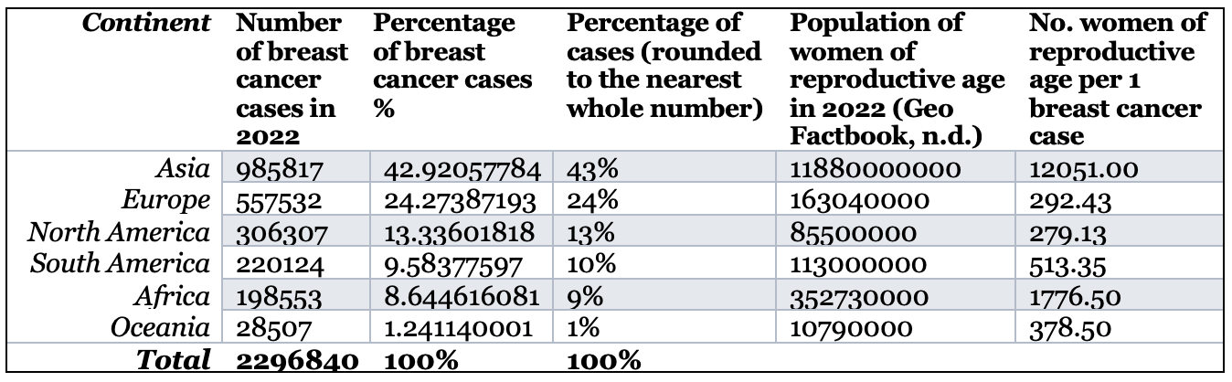
Table 1: Prevalence of breast cancer across the world. This table depicts the incidence of breast cancer cases in each continent and how common breast cancer is in the year 2022 (Freihat, 2025).
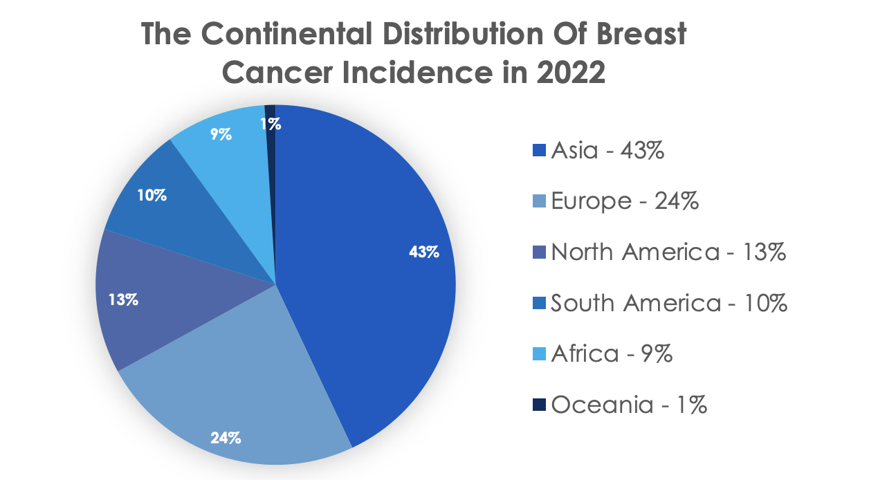
Figure 1: Prevalence of breast cancer across the world. A pie chart depicting the percentage of total breast cancer for each continent in 2022 (Freihat, 2025).
2.3 Aetiology and Risk Factors
There is no singular cause of breast cancer. However, there are numerous risk factors that increase the likelihood of developing breast cancer, some of which include:
- Presence of family history, which significantly increases risk due to the inheritance of key genes such as BRCA1, BRCA2 and PALB-2 (Lou, 2022). Studies have shown that women with a first-degree relative that has a prior breast cancer history have double the risk of getting breast cancer than women with no first-degree relations (Admoun, 2022).
- Sex, one of the most important risk factors for getting breast cancer: approximately 0.5-1% of breast cancer cases are male (World Health Organisation, 2026), making women about 100 times more likely to develop breast cancer than men (Fox Chase Cancer Centre, 2015).
- Age, an important risk factor: Women over 50 years consist of the majority of diagnoses. Breast cancer risk continues to increase after 50 years up until 70 years or older, reaching the greatest risk of 7.0% of developing breast cancer (Admoun, 2022).
- Geography, ethnicity and race, which can dramatically amplify the risk of breast cancer as demonstrated by North America, North and West Europe, Australia and New Zealand, where incidence rates are higher than the rest of the world (Admoun, 2022), such as in Europe for every one breast cancer case there are 292.43 people (see Table 1).
2.4 Types and Signs of Breast Cancer
Once breast cancer develops, it can appear in several pathological and molecular forms, each with variable clinical characteristics, that determine the treatment process for the patient. Histologically, it is broadly classified into non-invasive (in situ) cancers and invasive cancers (Tan et al., 2020). Non-invasive cancers occur when the cancer cells are still inside the milk ducts or lobules where they first formed and have not yet invaded the surrounding breast tissue. Such cancers typically include: ductal carcinoma in situ (DCIS) and lobular carcinoma in situ (LCIS) (Tan et al., 2020).
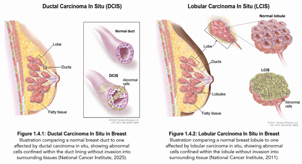
Figure 2: Ductal and lobular carcinoma in situ. These illustrations compare a healthy breast duct and lobule to one affected by carcinoma in situ (National Cancer Institute, 2011; 2025).
Beyond histological classification, breast cancer is also categorised at the molecular level, based on the presence of hormone receptors and HER2 protein expression, into three major subtypes: HER2 negative, HER2 positive and triple-negative (Waks & Winer, 2019).
Clinically, breast cancer usually presents itself as a painless breast lump. However, additional signs such as nipple inversion or discharge, skin dimpling, localised redness or thickening and breast asymmetry may indicate disease progression (Sharma & Saripilli, 2025). Since these also appear in benign breast conditions, accurate recognition and evaluation are essential for early detection and improved patient outcomes (Sharma & Saripilli, 2025).
2.5 Stages and Symptoms of Breast Cancer
Symptoms of breast cancer often become noticeable once the tumour has grown large enough to affect surrounding tissue. This means that early stage breast cancer often comes with no noticeable symptoms at all, thus requiring regular screening mammograms. Noticeable symptoms may include the sensation of a breast “lump” that is hard, irregular or painless. Irregularities surrounding the nipple’s discharge or skin lesions may also occur.
Following clinical presentation, breast cancer is staged to determine disease extent and guide treatment decisions. The stages of development of breast cancer are dictated using the standardised TNM (tumour, node, metastasis) system.
- Stage 0 breast cancer involves non-invasive cells that have not been found in nearby lymph nodes and have not metastasised to other organs.
- Stage 1 breast cancer is similarly limited to small tumours of 2cm or less.
- Stage 2 breast cancer covers tumours of up to 5cm in size. Both stage 1 and 2 are still limited in their spread towards nodes.
- Stage 3 breast cancer is differentiated from previous stages by its size (above 5cm) and potential to spread towards local nodes, being defined as a “locally advanced cancer”.
- Stage 4 breast cancer occurs when the tumour metastasises. Metastases are present in distant organs, with up to microscopic clusters surrounding auxiliary lymph nodes.
Stage 0-2 breast cancer often takes months to years to develop. It lasts proportionately longer than later stages due to the slow dividing nature of cells in the breast, allowing a large window of time to contain it.
2.6 Treatment of Breast Cancer
Breast cancer can be treated in multiple ways. Most commonly, the patient would undergo a lumpectomy, surgical removal of the lump, or a mastectomy which is the surgical removal of the breast (World Health Organisation, 2025).
With lumpectomy, a surgeon removes the cancerous tissue from the breast. The lymph nodes beside the breast may also be removed to minimise the chance for recurrence. The entire surgical process takes 1-2 hours, and 1-2 overnight stays may be necessary based on the situation (Cleveland Clinic, 2022). It is typically followed by radiation therapy to kill any remaining cancer cells left within the body, usually taking about 20 minutes per day and 5 days per week for 3-6 weeks (American Cancer Society, 2023). In a mastectomy, the breast and lymph nodes are removed, in which the cancer will then be assessed based on extent and spreading. Aesthetic surgeries such as breast reconstruction can also be done upon request (American Cancer Society, 2023).
Whether a lumpectomy or mastectomy is performed depends on the extent of spreading and location of the cancer. If the cancer is present under the nipple, the spreading is extensive or the patient does not wish to receive radiation therapy, mastectomy is often the preferred option (National Cancer Institute, 2025).
Early detection of breast cancer can significantly reduce its mortality. When the cancer is localised in the breast during surgery, the survival rate is 99%; when spread to the lymph nodes, the rate of survival falls to 86%, further falling to 30% when it spreads to other parts of the body (Cleveland Clinic, 2022). Through advances in treatment technology and increase in global awareness, breast cancer mortality in high income countries has dropped by 40% between 1980 and 2020. Many countries since then have achieved a mortality reduction of 2-4% per year (WHO, 2025).
3. Current Diagnosis of Breast Cancer
3.1 MRI and Biopsy
Magnetic resonance imaging is one of the most sensitive breast imaging modalities, particularly in women with dense breasts, those at high risk and when conventional imaging is unclear (Aristokli, 2022; Schoub, 2018; Mann, 2019; Chiarelli, 2020; Sardanelli, 2024). It helps define disease extent and can reveal additional lesions before treatment, improving surgical planning (Aristokli, 2022; Chiarelli, 2020; Sardanelli, 2024). However, its lower specificity means some findings may not be malignant, so MRI is best used as a complementary test rather than a replacement for pathology (Mann, 2019; Sardanelli, 2024).
On the other hand, biopsy provides the definitive pathological confirmation of whether a suspicious breast lesion is cancer (Silva, 2023; Zhang, 2013). Current guidelines recommend core needle biopsy as the preferred initial technique because it is less invasive while providing a tissue sample for testing (Silva, 2023; Sanderink, 2025). If a lesion can only be seen on MRI, an MRI-guided biopsy may be needed (Myers, 2015; Sanderink, 2025). This step is important because imaging can show a suspicious finding, but only a biopsy can determine whether it is cancer and help guide treatment (Myers, 2015; Silva, 2023).
3.2 Mammography and Overview of Imaging Techniques, Strengths and Limitations of Current Imaging Methods
Medical imaging plays a central role in the screening, detection and evaluation of breast cancer. Mammography is widely used for breast cancer screening, while digital breast tomosynthesis (DBT) may also be used depending on factors such as age, breast density, individual risk and clinical guidelines (Expert Panel on Breast Imaging et al., 2024). Evidence from randomised trials and clinical guidelines show that regular mammographic screening is linked to reduced risk of dying from breast cancer, which supports its continued use as the standard screening method (Marmot et al., 2013; Oeffinger et al., 2015). However, the effectiveness of mammography is influenced by breast density. Dense fibroglandular tissue can obscure breast lesions on mammograms, reducing mammographic sensitivity and increasing the risk of missed cancers (Yuan et al., 2020; Lee et al., 2024). Mammographic screening may also result in false-positive findings, recalls and further tests for women who do not actually have breast cancer (Oeffinger et al., 2015). These observations indicate that mammography offers significant clinical benefits, but does not work equally well for different patient groups.
When mammography alone does not provide sufficient diagnostic information, supplementary ultrasound is often used, particularly for women with dense breast tissue. Systematic reviews indicate that adding ultrasound to mammography can detect additional cancers that were not seen on the initial mammogram (Yuan et al., 2020; Vourtsis & Berg, 2019). Still, this higher cancer detection rate may come with lower specificity, more false-positive results and the need for more biopsies. For this reason, ultrasound should be viewed as a complementary tool rather than a direct substitute for mammography.
Overall, current research suggests that using multiple imaging techniques provides a more effective approach to breast cancer diagnosis as the choice of imaging depends on factors such as breast density, cancer risk and clinical presentation. Mammography remains the primary method for routine breast cancer screening, while ultrasound provides additional diagnostic information for selected patients. Ongoing challenges, including false-positive results, lower sensitivity in dense breasts and differences in how images are interpreted, have led to research on AI systems that could support radiologists by improving lesion detection and classification, assisting workflow management and supporting clinical decision-making (Lauritzen et al., 2024).
3.3 Clinical Evaluations, Staging and Tissue Diagnosis
Although imaging plays a central role in breast cancer detection, diagnosis also relies on clinical evaluation and pathological confirmations. Clinical evaluation begins with obtaining a detailed medical history, including the patient’s age, family history of breast or ovarian cancer, genetic risk factors such as BRCA1 and BRCA2 mutations, reproductive history, previous breast conditions and symptoms such as a breast lump, nipple discharge or changes in the skin (Bhushan et al., 2021; Feigin et al., 2006). A physical examination is then performed to evaluate the size, location and movement of the breast lump and to check whether nearby lymph nodes are enlarged or affected (Feigin et al., 2006).
A tissue diagnosis is essential to confirm breast cancer. This is done with a core needle biopsy in which the tissue sample is examined under a microscope to identify the type of breast cancer (Bhattacharyya et al., 2013). It is also tested for biomarkers, including estrogen receptor (ER), progesterone receptor (PR), HER2, Ki-67 proliferation index and tumour grade. These biomarkers help doctors determine how aggressive the cancer is and which treatments are most likely to be effective (Koh & Kim, 2018). The biopsy results must match the patient’s symptoms and clinical findings to ensure an accurate diagnosis. If the results are unclear or do not match the clinical evaluation, another biopsy or surgery may be needed to obtain more tissue (Bhattacharyya et al., 2013). In some patients, additional genetic tests called multigene expression assays are used to estimate the risk of cancer returning and determine whether chemotherapy is likely to be beneficial (Koh & Kim, 2018; Kubiak et al., 2026).
Once breast cancer is confirmed, the disease is staged using the American Joint Committee on Cancer (AJCC) staging system, a standard system used by doctors to describe how advanced a person’s cancer is. This system evaluates the size of the tumour, whether the cancer has spread to nearby lymph nodes and whether it has spread to other parts of the body. It also considers tumour grade and biomarker results to more accurately predict how the cancer might behave (Koh & Kim, 2018). By combining information from clinical evaluation, tissue analysis and tumour biology, doctors can create a personalised treatment plan and provide an accurate prediction of the prognosis (Kubiak et al., 2026). When used correctly, artificial intelligence can improve the accuracy of these evaluations by analysing large amounts of clinical and biological data, which will then help make better treatment decisions and more precise prognostic predictions.
4. Artificial Intelligence Models
4.1 Machine Learning
Machine learning is a subset of AI focused on building algorithms that learn patterns from data to make predictions without being programmed explicitly (Stranieri, 2022). By analysing mammograms to spot dangerous lesions that the naked eye would miss, calculating an individual’s risk of getting breast cancer and forecasting disease occurrence and treatment response, it plays a crucial role in breast cancer screening (Conner & Roldan, 2026).
The most popular algorithms include support vector machines (SVMs) and random forests (RF). By determining the ideal boundary between several classes, SVMs categorise tumours as benign or malignant, while RF integrates decision trees to increase predictive accuracy and decrease overfitting.
The use of AI in breast cancer cases has evolved from traditional machine learning algorithms to hybrid systems that combine machine learning with deep learning, enabling more accurate detection, diagnosis and risk prediction (Chaurasia et al., 2018). Early studies mainly evaluated algorithms such as SVMs and naïve Bayes, while newer research combines central neural networks (CNNs) with machine learning classifiers to improve feature extraction and prediction (Sherazi et al., 2020)
Machine learning is increasingly viewed as a clinical decision-support tool rather than a replacement (HIMSS, 2025). Across studies, AI is used to assist early detection and help prioritise suspicious cases, while final diagnosis remains clinician-led (AlOmair et al., 2026).
Despite high reported accuracies above 95%, researchers across studies acknowledge that limited dataset diversity remains a major barrier to clinical implementation (Chaurasia et al., 2018) as most studies rely on benchmark datasets, highlighting the need for validation using more diverse patient populations (Saharan et al., 2025).
4.2 Generative AI and Multimodal AI
Multimodal AI is shifting from unimodal imaging, which analyses only one data type, to intermediate fusion that enables feature-level cross-modal interaction and typically outperforms early or late fusion in heterogeneous settings (Zhang et al., 2026; Hassan et al., 2026). Intermediate fusion encodes each modality separately and then combines embeddings using attention, gating or graph-based modules, which helps handle missing or noisy modalities (Zhang et al., 2026; Hassan et al., 2026). The Framework for Exon-Specific Survival and Classification Analysis (FESCA) illustrates contrastive cross-modal semantic alignment between whole slide images (WSI), which are high-resolution digital pathology slides, and mRNA plus modality-adaptive fusion, reporting the area under the precision-recall curve (AUC-PR) as an imbalance-robust metric that reliably measures accuracy when positive cancer cases are rare in the dataset (Wang et al., 2026).
Generative AI is extending “detection” toward visual-language interaction and report-like outputs, yet mammogram large vision-language model (LVLM) performance, which integrates image and text processing, can be near random without domain adaptation (Hassan et al., 2026; Zhu et al., 2025). The Mammogram Visual Question-Answering dataset (MammoVQA) benchmarks 12 LVLMs and introduces LLaVA-Mammo, showing substantial gains after fine-tuning for BI-RADS-style tasks, which follow the standardised guidelines for breast imaging reporting and data systems (Zhu et al., 2025).
Generative models mitigate data scarcity and missing modalities via generative adversarial networks (GANs) or diffusion synthesis and reconstruction, which generate realistic synthetic patient data (Abdullakutty et al., 2024; Rofena et al., 2026). A cycle-consistent generative adversarial network (CycleGAN) can synthesise contrast-enhanced spectral mammography (CESM) from full-field digital mammography (FFDM) and still improve virtual-biopsy classification, a non-invasive computer method to predict tissue malignancy (Rofena et al., 2026). Finally, a score-based diffusion model reconstructs undersampled breast MRI with high radiologist ratings at acceleration factor 2, which effectively doubles the scanning acquisition speed (Okolie et al., 2025).
4.3 Deep Learning AI Models
Recent evidence demonstrates that AI systems for breast cancer increasingly use advanced machine learning and deep learning techniques to analyse different types of patient data. Modern AI models can integrate medical imaging, pathology and clinical information to provide more accurate and personalised predictions for diagnosis, prognosis and treatment planning (Jiang et al., 2024; Gao et al., 2025).
These findings show that several AI methods have been developed to support breast cancer care. Deep learning models, particularly CNNs, are widely used to analyse mammograms, MRI scans and digital pathology images, enabling the detection of subtle patterns that may be difficult for radiologists and pathologists to identify (Arunkumar & Sasikala, 2023; Jiang et al., 2024; Abdullah et al., 2025). Machine learning algorithms also combine imaging findings with genomic information to improve predictive accuracy, while radiogenomics integrates imaging features with genetic data to provide a better understanding of tumour biology (Qi et al., 2024; Gao et al., 2025). Furthermore, generative AI models, such as conditional generative adversarial networks (cGANs), can generate synthetic medical images and assist in predicting clinically relevant genetic alterations. Although machine learning models incorporating polygenic risk scores improve risk assessment by combining inherited genetic information with additional patient characteristics (Qi et al., 2024).
Overall, these studies suggest that AI performs best when multiple sources of patient data are integrated rather than relying on a single data type. By learning complex patterns from large datasets, these models have the potential to support more accurate and personalised clinical decision-making throughout breast cancer care. However, the reviews consistently emphasise that AI systems still require large, diverse, high-quality datasets. They also need external validation and prospective clinical testing before they can be safely and reliably implemented in routine healthcare practice (Jiang et al., 2024; Abdullah et al., 2025; Gao et al., 2025).
4.4 AI Medical Image Analysis
AI has transformed breast cancer image analysis by automating tumour detection, classification and segmentation, with recent advances in deep learning substantially improving diagnostic accuracy compared with conventional image analysis methods. For example, McKinney et al. (2020) trained an AI system to detect tumour presence from initial mammographic images using screening mammograms linked with biopsy-confirmed diagnoses.
Across studies, the general workflow of AI in imaging follows similar steps: data cleaning; detection of suspicious areas; segmentation using pixel-wise classification to locate specific tumour or lesion boundaries; and classification (Khosravi et al., 2025; Lamba, 2025; Suganyadevi et al., 2021). Although both traditional machine learning and deep learning follow similar preprocessing and classification pipelines, deep learning has become the more preferred approach because it can learn hierarchical image features directly from raw data. In contrast, traditional machine learning relies on handcrafted feature extraction, limiting its ability to capture the complex spatial patterns characteristic of mammographic images which leads to loss of vital information (Lamba, 2025).
While CNNs remain the most widely adopted class of deep learning due to their efficiency and strong performance in lesion segmentation from classification of high-resolution images, ViTs demonstrate improved ability to capture long-range spatial relationships by applying self-attention mechanisms. This may enable more accurate identification of subtle or diffuse imaging features that extend beyond the local receptive fields of CNNs. However, ViTs generally require substantially larger training datasets and greater computational resources, which may limit their clinical implementation compared with CNN-based models (Khosravi et al., 2025; Lamba, 2025; Pinto-Coelho et al., 2023).
A persistent challenge is the “black box” nature of many AI models as they often provide little insight into their decision-making processes, which makes it difficult for clinicians to verify whether predictions are based on clinically relevant imaging features, potentially increasing the risk of diagnostic errors. To address this, explainable AI (XAI) is used to clarify outputs for clinicians, improving trust and interpretability (Khosravi et al., 2025; Lamba, 2025).
Overall, while AI demonstrates strong potential in medical imaging, its effectiveness depends on robust dataset collection from diverse populations, advanced architectures such as CNNs and ViTs, and integration of algorithms like XAI to ensure transparency and reproducible outcomes.
5. Applications and Evidence
5.1 Impact of AI Implementation on Diagnostic Accuracy and Doctors’ Work
The real-world implementation of AI across international screening frameworks demonstrates its capacity to concurrently optimise clinical accuracy and administrative efficiency. For instance, Germany’s nationwide PRAIM implementation reported a higher cancer detection rate (CDR), which measures the number of cancers found per 1,000 screened women, with AI-supported double reading (6.7 vs 5.7 per 1,000) without worsening recall (Eisemann et al., 2025). In a paired noninferiority trial using partial autonomy, where an AI system triages low-risk scans and provides double-reading support only for higher-risk cases, radiologist workload fell by 63.6% while CDR increased (7.3 vs 6.3 per 1,000), though recall rose (Elías-Cabot et al., 2026). In Denmark, regional AI deployment shifted 66.9% of exams to single human reading, reducing workload by 33.5% while improving the recall rate, CDR and positive predictive value (PPV), which indicates the percentage of positive screening results that are confirmed to be actual cancer (Lauritzen et al., 2024). Prospective single-reading evidence from South Korea similarly shows CDR gains (5.70% vs 5.01%) without change in recall rate – the proportion of actual positive cases correctly identified by the model (Chang et al., 2025).
However, prospective deployment also introduces significant operational challenges and limitations that can undermine these clinical benefits. Multicentre feasibility work by Kelly et al. (2024) highlights that moving AI models from controlled environments to real-world clinics frequently triggers data distribution shifts – discrepancies that occur when the clinical data a model encounters in practice differs from the data it was trained on due to variations in patient demographics and imaging hardware. These shifts can lead to unpredictable algorithmic behaviour, requiring continuous monitoring and complex local threshold recalibration to prevent missed lesions or a surge in false positives. Furthermore, the authors emphasise that without rigorous multi-site validation, relying on AI risks exacerbating existing health inequities and compromising patient safety if the underlying system performs suboptimally on specific sub-populations.
5.2 Influence of Breast Density and Imaging Equipment on AI Performance
AI accuracy in breast cancer detection depends not only on algorithmic design but also on patient characteristics and imaging conditions (Le et al., 2019). Breast density, referring to how much of the breast is made up of glandular and fibrous (dense) tissue versus fatty tissue, is an important example of said characteristics that play a crucial role in AI performance (Murphy et al., 2025).
Breasts classified as dense are composed mainly of dense tissue, with relatively little fat. On mammographic images, dense tissue appears white while fat appears dark. This may be an issue since malignant tissue also appears white, leading to obscured tumours within dense regions. This reduces detection sensitivity in women with dense breasts, increasing the likelihood of missed or delayed diagnoses (Murphy et al., 2025).
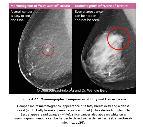
Figure 3: Mammographic comparison of fatty and dense tissue. Comparison of mammographic appearance of a fatty breast (left) and a dense breast (right). Fatty tissue appears dark while dense tissue appears white. Since cancer cells also appear white, tumours can be harder to detect within dense tissue (DenseBreast-info, Inc., 2026).
Studies evaluating AI-assisted mammography have shown that although AI can improve detection, its performance may vary across different breast densities, highlighting the need for models trained on diverse breast compositions (Rodríguez-Ruiz et al., 2019; Pesapane et al., 2026).
Additionally, variation in mammogram machines, scanning protocols and hospital practices can affect imaging quality and thus, AI performance (Le et al., 2019). Therefore, evaluations have shown that AI systems must undergo external validation across different scanners and healthcare settings to ensure consistent performance (Rodríguez-Ruiz et al., 2019).
5.3 Impact of Racial/Ethnic Differences on Accuracy of AI-based Mammogram Predictions
The accuracy of AI-based mammogram prediction models is influenced by racial and ethnic differences, mainly because many algorithms are developed using datasets that underrepresent minority populations. Despite increasing research into AI algorithms for mammogram analysis, most studies disproportionately feature white patients, with limited reporting of race or ethnicity. This lack of diversity contributes to flawed breast cancer predictions by AI models due to incomplete and unrepresentative datasets.
Multiple studies show that race and ethnicity influences AI predictions by leading to over- or under-estimation of breast cancer risk in non-Caucasian populations (Kerlikowske et al., 2025; Nguyen et al., 2024). Yet, their results diverge as Kerlikowske et al. (2025) demonstrate that Asian patients were overestimated for advanced breast cancer risk while Black patients were underestimated; in contrast, Nguyen et al. (2024) find Black patients more likely to receive false-positive scores and Asian patients less likely compared to white patients (odds ratio 1.5 and 0.7 respectively), reflecting the importance of further inclusion of ethnically diverse populations in breast cancer research. These discrepancies may be because of methodological differences: Nguyen et al. (2024) used a commercial image-analysis algorithm on true-negative mammograms, whereas Kerlikowske et al. (2025) applied a BCSC advanced breast cancer risk model predicting six-year advanced risk.
Both Miyawaki et al. (2025) and Amin et al. (2025) demonstrate that rapid growth in AI mammography research has not been accompanied by improvements in dataset diversity. Demonstrated in Figures 4 and 5 below, while the number of AI mammography studies have increased substantially between 2017 and 2023 with a 311% increase, reporting of participant race and ethnicity remain limited, with white patients representing an increasing proportion of study populations from 39% to 70%, although data for participants in 2017 is unavailable (Miyawaki et al., 2025). Similarly, Amin et al. (2025) identified major geographical disparities in breast imaging research, with far fewer studies conducted in Asia and none in Africa or Oceania compared with predominantly Caucasian populations. Collectively, these findings suggest that AI algorithms continue to be trained and evaluated using datasets that inadequately represent global populations.

Figure 4: Dataset diversity and mammography. This figure presents a line graph visualising the increase in the number of studies training AI algorithms with mammograms and the corresponding number of studies reporting race/ethnicity from 2017 to 2023 (Miyawaki et al., 2025).
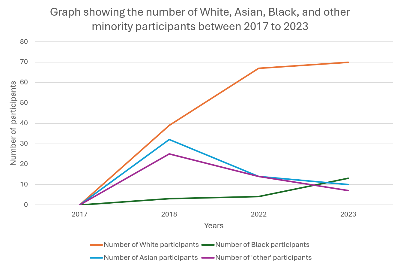
Figure 5: Minority participants in AI studies for breast cancer. This figure shows a line graph visualising the increase in the number of studies training AI algorithms with mammograms and the corresponding number of patients based on broad race categories from 2017 to 2023. Data in 2017 for the number of patients classified by race is unavailable (Miyawaki et al., 2025).
These gaps restrict generalisation of AI models for minority groups as they are trained with insufficient datasets and may lead to poor performance and inaccurate predictions for patients of said race or ethnicities. This aligns with Gichoya et al. (2023) as they argue that neglecting minority data perpetuates biased predictions across fields and that transparent documentation, diverse datasets and rigorous evaluation protocols should be enforced by researchers to ensure reliable outcomes.
Yet some studies are limited by the exclusion of non-English publications, leading to possibly overlooking research in minority populations, while generalising small groups (e.g., Pacific Islanders as “Others”) undermines accuracy (Kerlikowske et al., 2025). Together, these studies indicate that the primary challenge is not the AI models themselves but the representativeness of the datasets used to develop them. Until racially and ethnically diverse populations are routinely included in model development and external validation, AI-assisted mammography risks perpetuating existing healthcare inequalities, limiting both its clinical reliability and equitable implementation.
5.4 Application of AI for Histopathological Grading of Breast Cancer
In the domain of breast cancer, methods of histopathological grading continue to dominate its diagnosis and prognosis, with grading systems such as the Nottingham Histologic Grading (NHG) providing a standardised structure for the diagnosis of breast cancer aggressiveness to reveal treatment options. With the emergence of AI among various medical fields, deep learning AI models could reinforce the standardisation of histopathology grading by reconciling the inevitable inconsistencies associated with manual grading done by pathologists.
Specifically, AI models were best employed in assessment of parameters such as nuclear pleomorphism, mitotic count and tubule formation; by evaluating these three microscopic features, a pathological report for breast cancer aggressiveness can be formed. Typically, processes of this sort form the basis of the NHG system.
AI can also guide the development of treatment strategies and foresee outcomes such as treatment response, probability of survival and risks of recurrence. Furthermore, available multimodal datasets that provide AI with insight to realistic situations can be integrated for increased accuracy.
Over the recent years, numerous studies have concluded that the accuracy and reliability of AI-based methods have proven not far from those of expert pathologists. This concordance was demonstrated by a study conducted in 2022, where the grading of a deep learning system for the three areas of the NHG agreed with the judgement of three expert pathologists. Particularly, the AI model used in the study demonstrated excellence in detection of stage 0 breast cancer ductal carcinoma in situ with a near perfect area under the curve (AUC) of 0.98. Specificity and sensitivity of the system was also scored above 93% in various carcinoma detection.
Overall, harnessing AI as a tool in the processes of histopathological grading reduces variability and introduces higher efficiency in the diagnosis and prognosis of breast cancer. Nonetheless, challenges persist in establishing trust and reliability for further commercial applications.
5.5 Evidence for AI in Breast Cancer Genetics and Genomics
Recent evidence demonstrates that the integration of AI has improved breast cancer screening, diagnosis, prognosis and treatment selection while supporting more personalised patient care (Lauritzen et al., 2023; Lai et al., 2024; Huang et al., 2024; Miao et al., 2026). Across recent studies, AI has been successfully applied to multiple aspects of breast cancer management, including risk prediction, cancer detection, diagnosis and the identification of clinically relevant genetic mutations. AI systems have also been developed using a variety of data sources, such as mammographic and MRI images, digital pathology slides, genomic information (DNA information), transcriptomic data and clinical patient records (Lauritzen et al., 2023; Lai et al., 2024; Miao et al., 2026).
These findings show that multiple types of patient information together produce predictions that are more accurate than the use of a single data source (Lai et al., 2024; Huang et al., 2024; Miao et al., 2026). By combining medical imaging and genomic information, AI models can predict more accurately if cancer will spread to the lymph nodes, as well as how the patient will respond to treatment before surgery (Lai et al., 2024). This can help doctors determine which treatments will likely be most effective and choose targeted therapies for the individual. The AI models can look at pathology images and genetic data to find mutations that are important for patient care (Miao et al., 2026). Through these findings it shows that screening can be personalised for each patient.
Overall, the reviewed studies agree that AI has the potential to improve breast cancer care by supporting diagnosis, risk prediction and personalised treatment planning. However, they all differ in how the technology is evaluated: Schaffter et al. (2025) and Kim et al. (2025) evaluate AI in real-world clinical breast cancer screening programmes. In contrast, Wu et al. (2024), Lauritzen et al. (2023), Lai et al. (2024), Huang et al. (2024), and Miao et al. (2026) focus on developing, validating or reviewing AI models using existing datasets rather than implementing them in routine clinical practice.
6. Challenges and Ethical Considerations
6.1 Data Quality, Availability and Generalisation
The accuracy of AI models in healthcare must be based on and developed using diverse, wide-set data that represents varied populations (Bergman et al., 2023). Diversity in training datasets is essential as larger, more varied datasets allow the models to recognise patterns more accurately. Using smaller datasets, particularly in medical imaging, limits the model’s ability to recognise rare diseases and interpret imaging accurately.
Class imbalance is another key factor to consider. This occurs when one class of data has more samples than the other, biasing the model towards the majority class. When considering class imbalance in AI-assisted medical imaging, this could mean common diseases are diagnosed more reliably while rarer conditions are more likely to be missed (Albattah & Khan, 2025). AI models also tend to lose accuracy when used in a different hospital than the one they were originally trained in. This shift may be caused by the patient demographic or the hospital’s resources (Singh, Mhasawade & Chunara, 2022). This phenomenon is known as a domain shift (Musa, Prasad & Hernandez, 2025).
To address this, models should be trained on both multicentre and multinational datasets, combining data across hospitals in different countries and regions to understand how treatment may differ depending on age or health conditions and allow generalisability across the field (Rockenschaub et al., 2024). This allows AI to perform across diverse healthcare settings, ultimately improving patient safety and reliability (Singh, Mhasawade & Chunara, 2022).
6.2 Bias, Equity and Accountability
AI deployment in breast cancer screening risks systemic disparities if dataset, selection and algorithmic biases remain unaddressed. AI models trained on localised datasets fail to generalise across diverse clinical environments, equipment manufacturers or patient populations. 12 state-of-the-art general and medical LVLMs performed statistically equivalent to random guessing on the multicenter MammoVQA benchmark (Zhu et al., 2025). This illustrates that pre-training on basic medical/web data fails to capture varied mammographic traits across broad populations. Most public and institutional mammography datasets (e.g., CBIS-DDSm, Ibreast) predominantly represent populations of European descent. AI models trained without adequate racial diversity misclassify subtle abnormal findings in skin or tissue densities that vary across ancestral populations (Magni et al., 2022). Mitigating these diagnostic inequities requires training models on diverse, multi-institutional datasets (e.g., MammoVQA), applying techniques like SMOTE to prioritise recall and conducting independent external validation. Crucially, legal and clinical liability remains with the radiologist, reinforcing AI as a supportive tool within human-centric care.
6.3 Transparency, Explainability and Trust
Deep learning architectures often function as black boxes, delivering diagnostic metrics without clinical rationale (Saharan et al., 2025). Clinicians cannot defend therapy recommendations or invasive biopsies based on an inexplicable black box output since they operate under medical-legal liability and evidence-based medicine. To bridge this trust gap, explainable AI is essential; methods such as the Shapley Additive Explanations (SHAP) approach measure individual feature contributions, while visual questional answering models provide interactive diagnostic reasoning (Ghauth, 2026).
Ultimately, trust in AI is built through accountability rather than automation. Instead of taking the place of clinical expertise, AI should serve as a decision-supporting tool. Human oversight remains essential to identify errors, communicate uncertainty to patients and ensure that diagnostic decisions consider both algorithmic outputs and individual clinical context.
6.4 Ethical, Legal and Governance Challenges
The use of AI in breast cancer care relies on large and high-quality datasets, which is why it is essential to have proper privacy measures and data governance if the technology is to be used in a safe and ethical manner (Davenport & Kalakota, 2019; Carter et al., 2020; World Health Organisation, 2021). To protect patient privacy, it is necessary to ensure secure data sharing, obtain informed consent, establish clear rules about data ownership and have effective cybersecurity to minimise the risk of unauthorised access and the misuse of health information (Carter et al., 2020; World Health Organisation, 2021). Current legal frameworks, including the General Data Protection Regulation (GDPR) in the European Union and the Health Insurance Portability and Accountability Act (HIPAA) in the United States, offer regulatory safeguards for the collection, processing and sharing of patients’ health data within their respective jurisdictions (Carter et al., 2020; World Health Organisation, 2021).
As AI becomes incorporated into clinical care, clear accountability for AI-assisted clinical decisions will be essential, particularly when errors occur or when continuously learning systems change their performance over time.
Moreover, unequal access to AI-enabled healthcare and the underrepresentation of certain populations in training datasets could increase the existing disparities in breast cancer care, highlighting the need for governance frameworks that promote equitable access and ensure that AI systems perform reliably across diverse populations (Li, 2022; Bayard, 2022; World Health Organisation, 2021).
6.5 Implementation Challenges
AI technology has the potential to correct human error, but successfully integrating it into practice relies on adapting these models to existing clinical workflows and the needs of healthcare workers. Rather than implementing AI wherever possible, healthcare systems should target areas where it can improve efficiency. Successful implementation also requires adequate technical infrastructure, including reliable digital systems and sufficient computing power. Equally important, healthcare professionals need to build the digital and AI literacy necessary to understand and use these technologies effectively (Bajwa et al., 2021)
AI must be clinically validated before widespread adoption to ensure it is both safe and effective. Testing should take place in real clinical environments using data from hospitals in different cities or countries. To achieve clinical validation, hospitals should: first assess the accuracy and technical performance of the AI; then, evaluate its performance with real patients; and finally, determine its utility by whether it improves patients’ diagnoses. Accuracy alone is not sufficient enough for the models to be implemented widely. The models must also be tested in live healthcare settings and evaluated for safety and to what extent it really helps patients and providers. One established method for evaluating clinical utility is through RCTs (Park, Choi & Byeon, 2021). In this context, an RCT would compare the AI assisted care to standard care without AI (Hariton & Locascio, 2018).
While AI can improve healthcare efficiency, it raises environmental and financial concerns. This is because it requires significant computing power, energy and resources (Bignami et al., 2025) One proposed solution to this is green AI. Green AI focuses on integrating renewable energy and optimising energy consuption (UST, n.d.). Additionally, hospitals must invest in new technology, data infrastructure, staff training and maintenance (Bignami et al., 2025). For AI to be fully integrated into clinical practice, healthcare systems must ensure that the benefits of using AI are greater than the costs of purchasing and maintaining it.
6.6 Women’s Perspectives and Global Acceptance of AI in Breast Cancer Care
Women’s trust and acceptance are essential for implementing AI in breast cancer care. Available evidence suggests that many women are more willing to accept AI in breast cancer care when it supports, rather than replaces, healthcare professionals, with surveys from Norway and Italy consistently showing a strong preference for physician involvement in AI-assisted mammogram assessment (58.5% preferred AI-assisted mammogram assessment in Norway; 94% believed radiologists should always examine mammograms in Italy) (Carter et al., 2024; Holen et al., 2024; Pesapane et al., 2023).
Acceptance is influenced by concerns about privacy, bias, accountability, reduced human interaction and professional de-skilling, as well as by factors like AI knowledge, digital literacy, education and health literacy (Lennox-Chhugani et al., 2021; Carter et al., 2023, 2024; Hemphill et al., 2023; Elhadi et al., 2025).
Across studies conducted in Norway, Italy, Sweden, Australia, the United Arab Emirates and the United States, a broadly consistent preference has been reported for AI to be used as a clinical decision-support tool under the supervision of a healthcare professional, rather than as an independent decision-maker (Pesapane et al., 2023; Carter et al., 2024; Holen et al., 2024; Johansson et al., 2024; Elhadi et al., 2025; Ozcan et al., 2025).
Nevertheless, the available evidence is drawn from high-income countries, highlighting the need for further research in low- and middle-income settings, where differences in healthcare infrastructure, access to technology, digital literacy and cultural attitudes may influence the acceptance and implementation of AI in breast cancer care (Carter et al., 2024).
6.7 Future Perspectives: Toward Trustworthy and Patient-Centred AI
In a 2025 multi-modal study on AI training and its effects on performance in detecting breast cancers from mammograms, an initial model trained on approximately 500,000 mammography exams, obtained an area under the receiving operator characteristic curve (AUROC) of 0.945. However after further training on more than 750,000 exams, its remaining error rate was reduced by 18.9-56.6%. (Park et al., 2025). Today, training continues with further advanced AI models, and if the previous study is extrapolated, AI models in the near future may reach exponentially improved levels of accuracy that parallel high-skill radiologists, providing strong evidence for a trustworthy system of identifying breast cancers.
However, breast cancer differs across various ethnicities across the world. To create a truly patient-centred AI system, models must be trained to be effective at identifying breast cancers in all ethnicities. Currently, world-leading training datasets like EMBED often lack representation for certain Asian populations, thus creating uncertainty when used in patients of ethnicities outside of the dataset. For example, the 2025 multi-modal study utilised a training population which was drawn mainly from US healthcare systems, meaning its performance may not reflect evenly across all ethnic groups worldwide. This serves as a major bottleneck for the world-wide implementation of AI in detecting breast cancers, which must be confronted in future datasets through the equal representation of all ethnicities in training.
7. Conclusion
Breast cancer is the leading cause of cancer-related death in women. However, it has a high recovery rate if caught early and treatment is started before the cancer becomes too advanced. Similarly, current diagnoses have flaws due to the presence of human clinicians, which leads to possible inaccuracies due to fatigue, lack of concentration and, sometimes, lack of experience. These flaws can possibly be resolved by introducing AI into the diagnosis process and helping reduce missed cancers.
Current breast cancer diagnoses depend on combining clinical evaluations, imaging, biopsy and staging to ensure accurate and personalised care. Each method has strengths and limitations, but together they help doctors make accurate diagnoses and treatment decisions. By addressing these limitations, AI has the potential to improve diagnostic accuracy further.
AI is transforming breast cancer care by improving early detection, diagnosis, risk prediction and personalised treatment planning. Machine learning, deep learning, multimodal AI, generative AI and advanced medical image analysis each contribute unique strengths, while their integration offers the greatest clinical potential. Despite high reported accuracy across many studies, challenges such as limited dataset diversity, lack of external validation, interpretability and regulatory considerations continue to restrict routine clinical implementation. In order to guarantee the safe, reliable and equitable integration of AI systems into clinical practice, future research should prioritise robust validation, diverse patient populations and explainable AI systems.
The use of AI has demonstrated that there is potential to improve breast cancer screening, diagnosis, histopathological grading and personalised risk assessment across diverse clinical settings. Developments in multimodal AI, medical image analysis and genomics have enhanced diagnostic accuracy and supported more efficient clinical workflows. However, the reviewed evidence also highlights important challenges, including performance differences across breast density, imaging equipment and racial and ethnic groups, as well as the need for greater transparency and external validation. Future research should prioritise diverse datasets, explainable AI and robust clinical evaluation to ensure equitable and reliable implementation in routine practice.
AI is expected to improve breast cancer diagnosis through greater accuracy and efficiency, but its future depends on addressing bias, ensuring transparency, protecting patient privacy and ensuring AI performs well across diverse patient populations. Rather than replacing radiologists, AI should support clinical decision-making, promote fair and inclusive care, build trust and improve patient care across diverse populations.
Bibliography
Abeelh, E.A. & AbuAbeileh, Z. (2024) ‘Comparative effectiveness of mammography, ultrasound, and MRI in the detection of breast carcinoma in dense breast tissue: A systematic review’, Cureus, 16(4), e59054.
Abdullah, K.A., Marziali, S., Nanaa, M., Sanchez, L.E., Payne, N.R. & Gilbert, F.J. (2025) ‘Deep learning-based breast cancer diagnosis in breast MRI: systematic review and meta-analysis’, European Radiology, 35(8), pp. 4474-4489.
Abdullakutty, F., Akbari, Y., Al-Maadeed, S., et al. (2024) ‘Histopathology in focus: a review on explainable multi-modal approaches for breast cancer diagnosis’, Frontiers in Medicine, 11.
Admoun, C. & Mayrovitz, H.N. (2022) ‘The etiology of breast cancer’, in Mayrovitz, H.N. (ed.) Breast Cancer (Brisbane, AU: Exon Publications).
Ahn, J.S., Shin, S., Yang, S.-A., Park, E.K., Kim, K.H. & Cho, S. I. (2023) ‘Artificial intelligence in breast cancer diagnosis and personalized medicine’, Journal of Breast Cancer, 26(5), pp. 405-435.
Albattah, W. & Khan, R.U. (2025) ‘Impact of imbalanced features on large datasets’, Frontiers in Big Data, 8, 1455442.
AlOmair, O., Zakaria, O.M., Alabdullatif, M. & Khurshid, Z, (2026) ‘Optimized KNN with domain-informed features and LIME explainability for improved breast cancer classification’, BMC Medical Informatics and Decision Making, 26(1), 138.
American Cancer Society (2026) ‘Treating Breast Cancer’, ACS [online]. <https://www.cancer.org/cancer/types/breast-cancer/treatment.html>
Amin, A., Acharya, D., Koteshwara, P. et al. (2025) ‘A systematic literature review on mammography: deep learning techniques for breast cancer detection with global and Asian perspectives’, BMC Cancer, 25(1), 1627.
Aristokli, N., Polycarpou, I., Themistocleous, S.C., Sophocleous, D. & Mamais, I. (2022) ‘Comparison of the diagnostic performance of magnetic resonance imaging (MRI), ultrasound and mammography for detection of breast cancer based on tumour type, breast density and patient history: A review’, Radiography, 28(3), pp. 848-856.
Bahadir, C.D., Omar, M., Rosenthal, J. et al. (2024) ‘Artificial intelligence applications in histopathology’, Nature Reviews Electrical Engineering, 1(2), pp. 93-108.
Bajwa, J., Munir, U., Nori, A. & Williams, B. (2021) ‘Artificial intelligence in healthcare: transforming the practice of medicine’, Future Healthcare Journal, 8(2), e188-e194.
Bayard, S., Fasano, G., Gillot, T. et al. (2022) ‘Breast cancer disparities and the digital divide’, Current Breast Cancer Reports, 14(4), pp. 205-212
Bergman, A.S., Hendricks, L.A., Rauh, M. et al. (2023) ‘Representation in ai evaluations’, 2023 ACM Conference on Fairness, Accountability, and Transparency, Chicago IL USA: ACM, pp. 519-533.
Bernardi, F.A., Alves, D., Crepaldi, N. et al. (2023) ‘Data quality in health research: integrative literature review’, Journal of Medical Internet Research, 25, e41446.
Bhattacharyya, D. (2013) ‘Diagnosis of breast cancer by tissue analysis’, Chinese Journal of Cancer Research, 25(1), pp. 39-43.
Bhushan, A., Gonsalves, A. & Menon, J.U. (2021) ‘Current state of breast cancer diagnosis, treatment, and theranostics’, Pharmaceutics, 13(5), 723.
Bignami, E., Darhour, L.J. & Bellini, V. (2025) ‘Sustainable AI in medicine: navigating innovation, challenges, and environmental impact’, Health Economics Review, 15, 110.
Carriero, A., Groenhoff, L., Vologina, E., Basile, P. & Albera, M. (2024) ‘Deep learning in breast cancer imaging: State of the art and recent advancements in early 2024’, Diagnostics, 14(8), 848.
Carter, S.M., Carolan, L., et al. (2023) ‘Australian women’s judgements about using artificial intelligence to read mammograms in breast cancer screening’, Digital Health, 9, 20552076231191057.
Carter, S.M., Rogers, W., Win, K.T., et al. (2019) ‘The ethical, legal and social implications of using artificial intelligence systems in breast cancer care’, The Breast : official journal of the European Society of Mastology, 49, pp. 25-32.
Carter, S.M., Popic, D., Marinovich, M.L., et al. (2024) ‘Women’s views on using artificial intelligence in breast cancer screening: A review and qualitative study to guide breast screening services’, The Breast, 77, 103783.
Chang, Y.-W., Ryu, J.K., An, J.K., et al. (2025) ‘Artificial intelligence for breast cancer screening in mammography (Ai-stream): preliminary analysis of a prospective multicenter cohort study’, Nature Communications, 16(1), 2248.
Chaurasia, V. & Pal, S. (2018) ‘Prediction of Benign and Malignant Breast Cancer Using Data Mining Techniques’, SSRN Electronic Journal.
Chiarelli, A.M., Blackmore, K.M., Muradali, D. et al. (2019) ‘Performance measures of magnetic resonance imaging plus mammography in the High Risk Ontario Breast Screening Program’, JNCI: Journal of the National Cancer Institute, 112(2), pp. 136-144.
Cleveland Clinic (2023) ‘Breast Cancer Surgery’, Cleveland Clinic [online]. <https://my.clevelandclinic.org/health/treatments/8338-breast-cancer-surgery>
Conner, K. & Roldan, B. (2026) ‘Using AI to Detect Breast Cancer: What we know’, Breastcancer.org [online]. <https://www.breastcancer.org/screening-testing/artificial-intelligence>
Davenport, T. & Kalakota, R. (2019) ‘The potential for artificial intelligence in healthcare’, Future Healthcare Journal, 6(2), pp. 94-98.
DenseBreast-Info (n.d.) ‘Why does breast density matter on my mammogram?’, DenseBreast-Info [online]. <https://densebreast-info.org/patient-faqs/why-does-breast-density-matter-on-my-mammogram/>
Eisemann, N., Bunk, S., Mukama, T., et al. (2025) ‘Nationwide real-world implementation of AI for cancer detection in population-based mammography screening’, Nature Medicine, 31(3), pp. 917-924.
Elhadi, Y.A.M., Abdullahi, A.S., Shamsi, A.A. et al. (2025) ‘Women’s perspectives on integrating artificial intelligence in breast cancer screening services in Abu Dhabi, united Arab Emirates’, Scientific Reports, 16, 1209.
Elhakim, M.T., Stougaard, S.W., Graumann, O., Nielsen, M., Gerke, O., Larsen, L.B. & Rasmussen, B.S.B. (2024) ‘AI-integrated screening to replace double reading of mammograms: A population-wide accuracy and feasibility study’, Radiology: Artificial Intelligence, 6(6), e230529.
Elías-Cabot, E., Romero-Martin, S., Raya-Povedano, J.L., Rodriguez-Ruiz, A. & Alvarez-Benito, M. (2026) ‘AI-based triage and decision support in mammography and digital tomosynthesis for breast cancer screening: a paired, noninferiority trial’, Nature Medicine, 32(4), pp. 1296-1305.
Expert Panel on Breast Imaging et al. (2024) ‘ACR Appropriateness Criteria® female breast cancer screening: 2023 update’, Journal of the American College of Radiology, 21(6), S126-S143.
Feigin, K.N., Keating, D.M., Telford, P.M. & Cohen, M.A. (2006) ‘Clinical breast examination in a comprehensive breast cancer screening program: Contribution and cost’, Radiology, 240(3), pp. 650-655.
Fox Chase Cancer Center (2015) ‘Breast cancer inherited risk’, Fox Chase Cancer Center [online]. <https://www.foxchase.org/cancer-care-services/prevention-screening/risk-assessment/inherited-cancer/breast-cancer-inherited>
Freihat, O., Sipos, D. & Kovacs, A. (2025) ‘Global burden and projections of breast cancer incidence and mortality to 2050: A comprehensive analysis of GLOBOCAN data’, Frontiers in Public Health, 13, 1622954.
Gao, S., Liu, J., Li, L. et al. (2026) ‘Application of deep learning technology in breast cancer: a systematic review of segmentation, detection, and classification approaches’, Biomedical Engineering Online, 25(1), 19.
Geo Factbook (2025) ‘Female population of reproductive age 2022’, Geo Factbook [online]. <https://geofactbook.com/fact/female-population-of-reproductive-age/2022>
Ghauth, K.I. & Kustiawan, Y.A. (2026) ‘Explainable Artificial Intelligence (XAI) for Cancer Classification in Medical Imaging: A Systematic Review’, Machine learning & knowledge extraction, 8(5), 134.
Gichoya, J.W., Thomas, K., Celi, L.A. et al. (2023) ‘AI pitfalls and what not to do: mitigating bias in AI’, British Journal of Radiology, 96(1150).
Gradishar, W.J., Moran, M.S., Abraham, J., et al. (2024) ‘Breast cancer, version 3.2024, NCCN Clinical Practice Guidelines in Oncology’, Journal of the National Comprehensive Cancer Network, 22(5), pp. 331-357.
Ha, S.M., Jang, M.-J., Youn, I., Yoen, H., Ji, H., Lee, S.H., Yi, A. Chang, J.M. (2024) ‘Screening outcomes of mammography with AI in dense breasts: A comparative study with supplemental screening ultrasound’, Radiology, 312(1), e233391.
Hanahan, D. (2022) ‘Hallmarks of cancer: New dimensions’, Cancer Discovery, 12(1), pp. 31-46.
Hariton, E. & Locascio, J.J. (2018) ‘Randomised controlled trials—the gold standard for effectiveness research’, BJOG : an international journal of obstetrics and gynaecology, 125(13), 1716.
Hassan, M.M., Tahsin, A., Alam, G.R. et al. (2025) ‘Explainable multimodal fusion for breast carcinoma diagnosis: A systematic review, open problems, and future directions’, Computers in Biology and Medicine, 274.
Hemphill, S., Jackson, K., Bradley, S. & Bhartia, B. (2023) ‘The implementation of artificial intelligence in radiology: a narrative review of patient perspectives’, Future Healthcare Journal, 10(1), pp. 63-68.
HIMSS (2025) ‘How AI is reshaping clinical decision-making’, HIMSS Global Health Conference & Exhibition [online]. <https://www.himssconference.com/blog/how-ai-is-reshaping-clinical-decision-making-in-2025/>
Holen, Å.S., Martiniussen, M.A., Bergan, M.B., et al. (2024) ‘Women’s attitudes and perspectives on the use of artificial intelligence in the assessment of screening mammograms’, European Journal of Radiology, 175, 111431.
Huang, Z.H., Chen, L., Sun, Y., Liu, Q. & Hu, P. (2024) ‘Conditional generative adversarial network driven radiomic prediction of mutation status based on magnetic resonance imaging of breast cancer’, Journal of Translational Medicine, 22(1), 226.
Jiang, B., Bao, L., He, S., Chen, X., Jin, Z. & Ye, Y. (2024) ‘Deep learning applications in breast cancer histopathological imaging: diagnosis, treatment, and prognosis’, Breast cancer research: BCR, 26(1), 137.
Kelly, C.J., Wilson, M., Warren, L.M., et al. (2026) ‘Diagnostic accuracy, fairness and clinical implementation of AI for breast cancer screening: results of multicenter retrospective and prospective technical feasibility studies’, Nature Cancer, 7(3), pp. 494-506.
Kerlikowske, K., Chen, S., Sprague, B.L., Tice, J.A., Miglioretti, D.L., & Hubbard, R.A. (2025) ‘Effect of race and ethnicity on advanced breast cancer risk prediction model performance’, npj Digital Medicine, 8(1), 771.
Khosravi, P., Fuchs, T.J. & Ho, D.J. (2025) ‘Artificial intelligence-driven cancer diagnostics: enhancing radiology and pathology through reproducibility, explainability, and multimodality’, Cancer Research, 85(13), pp. 2356-2367.
Koh, J. & Kim, M.J. (2019) ‘Introduction of a new staging system of breast cancer for radiologists: An emphasis on the prognostic stage’, Korean Journal of Radiology, 20(1), pp. 69-82.
Kubiak, K., Bidzinska, J., Bednarek, M. & Szurowska, E. (2026) ‘Advances in breast cancer diagnostics: From screening to precision medicine’, Diagnostics, 16(8), 1181.
Kumar, S.A. & Sasikala, S. (2023) ‘Review on deep learning-based cad systems for breast cancer diagnosis’, Technology in Cancer Research & Treatment, 22.
Lai, J., Chen, Z., Liu, J., Zhu, C., Huang, H., Yi, Y., Cai, G. & Liao, N. (2024) ‘A radiogenomic multimodal and whole-transcriptome sequencing for preoperative prediction of axillary lymph node metastasis and drug therapeutic response in breast cancer: a retrospective, machine learning and international multicohort study’, International Journal of Surgery (London, England), 110(4), pp. 2162-2177.
Lamba, N. (2025) ‘Advances in AI for Medical Imaging: A Review of Machine and Deep Learning in Disease Detection’, Procedia Computer Science, 260, pp. 262-273.
Lauritzen, A.D., Lillholm, M., Nielsen, M., Karssemeijer, N. & Vejborg, I. (2024) ‘Early indicators of the impact of using AI in mammography screening for breast cancer’, Radiology, 311(3), e232479.
Lauritzen, A.D., von Euler-Chelpin, M.C., Lynge, E., et al. (2023) ‘Assessing breast cancer risk by combining ai for lesion detection and mammographic texture’, Radiology, 308(2), e230227.
Le, E.P.V., Wang, Y., Huang, Y., Hickman, S. & Gilbert, F.J. (2019) ‘Artificial intelligence in breast imaging’, Clinical Radiology, 74(5), pp. 357-366.
Lennox-Chhugani, N., Chen, Y., Pearson, V., et al. (2021) ‘Women’s attitudes to the use of AI image readers: a case study from a national breast screening programme’, BMJ Health & Care Informatics, 28(1), e100293.
Li, H., Whitney, H.M., Edwards, A. et al. (2022) ‘Impact of continuous learning on diagnostic breast MRI AI: evaluation on an independent clinical dataset’, Journal of Medical Imaging, 9(3), 034502.
Luo, J., Craver, A., Moore, K., Stepniak, L., King, J., Herbert, J. & Aschebrook-Kilfoy, B. (2022) ‘Etiology of breast cancer: A perspective from epidemiologic studies’, Journal of the National Cancer Center, 2(4), pp. 195-197.
Magni, V., Interlenghi, M., Cozzi, A. et al. (2022) ‘Development and Validation of an AI-driven Mammographic Breast Density Classification Tool Based on Radiologist Consensus’, Radiol Artif Intell, 4(2), e210199.
Makki, J. (2015) ‘Diversity of breast carcinoma: Histological subtypes and clinical relevance’, Clinical Medicine Insights: Pathology, 8, pp. 23-31.
Mann, R.M., Kuhl, C.K. & Moy, L. (2019) ‘Contrast-enhanced MRI for breast cancer screening’, Journal of Magnetic Resonance Imaging, 50(2), pp. 377-390.
Marmot, M.G., Altman, D.G., Cameron, D.A., Dewar, J.A., Thompson, S.G. & Wilcox, M. (2013) ‘The benefits and harms of breast cancer screening: An independent review’, British Journal of Cancer, 108(11), pp. 2205-2240.
Mayo Clinic (2025) ‘Breast cancer: Symptoms and causes’, Mayo Clinic [online]. <https://www.mayoclinic.org/diseases-conditions/breast-cancer/symptoms-causes/syc-20352470>
McKinney, S.M., Sieniek, M., Godbole, V. et al. (2020) ‘International evaluation of an AI system for breast cancer screening’, Nature, 577(7788), pp. 89-94.
Miao, J., Liu, Q., Zhao, J. et al. (2026) ‘Multimodal artificial intelligence predicts PIK3CA mutation in breast cancer from digital pathology and clinical data: a multicenter study’, Cancer Biology & Medicine, 23(3), pp. 430-450.
Miyawaki, I.A., Banerjee, I., Batalini, F., et al. (2025) ‘Global disparities in artificial intelligence-based mammogram interpretation for breast cancer: A scientometric analysis of representation, trends, and equity’, EJC, 220, 115394.
Murphy, P.C., McEntee, M., Maher, M. et al. (2025) ‘Assessment of breast composition in MRI using artificial intelligence – A systematic review’, Radiography, 31(2).
Musa, A., Prasad, R. & Hernandez, M. (2025) ‘Addressing cross-population domain shift in chest X-ray classification through supervised adversarial domain adaptation’, Scientific Reports, 15, 11383.
Myers, K.S., Kamel, I.R. & Macura, K.J. (2015) ‘MRI-guided breast biopsy: Outcomes and effect on patient management’, Clinical Breast Cancer, 15(2), pp. 143-152.
National Cancer Institute (2011) ‘Invasive ductal carcinoma’, NCI [online]. <https://www.cancer.gov/publications/dictionaries/cancer-terms/def/invasive-ductal-carcinoma>
National Cancer Institute (2011) ‘Invasive lobular carcinoma’, NCI [online]. <https://www.cancer.gov/publications/dictionaries/cancer-terms/def/invasive-lobular-carcinoma>
National Cancer Institute (2011) ‘Lobular Carcinoma in Situ (LCIS), NCI [online]. <https://www.cancer.gov/publications/dictionaries/cancer-terms/def/lcis>
National Cancer Institute (2025) ‘Ductal Carcinoma in Situ (DCIS), NCI [online]. <https://www.cancer.gov/types/breast/breast-cancer-types/dcis>
National Cancer Institute (2025) ‘Lumpectomy vs. Mastectomy’, NCI [online]. <https://www.cancer.gov/types/breast/treatment/surgery/choosing-lumpectomy-mastectomy>
National Health Service (2024) ‘Symptoms of breast cancer in women’, NHS [online]. <https://www.nhs.uk/conditions/breast-cancer-in-women/symptoms-of-breast-cancer-in-women/>
Nguyen, D.L., Ren, Y., Jones, T.M. et al. (2024) ‘Patient characteristics impact performance of ai algorithm in interpreting negative screening digital breast tomosynthesis studies’, Radiology, 311(2), e232286.
Oeffinger, K.C., Fontham, E.T.H., Etzioni, R., et al. (2015) ‘Breast cancer screening for women at average risk: 2015 guideline update from the American Cancer Society’, JAMA, 314(15), pp. 1599-1614.
Okolie, A., Dirrichs, T., Huck, L.C. et al. (2025) ‘Accelerating breast MRI acquisition with generative AI models’, European Radiology, 35(2), pp. 1092-1100.
Ozcan, B.B., Dogan, B.E., Xi, Y., Knippa, E.E. (2025) ‘Patient perception of artificial intelligence use in interpretation of screening mammograms: a survey study’, Radiology. Imaging Cancer, 7(3), e240290.
Park, J., Witowski, J., Xu, Y. et al. (2025) ‘A multi-modal ai system for screening mammography: integrating 2d and 3d imaging to improve breast cancer detection in a prospective clinical study’. arXiv, 2504.05636.
Park, S.H., Choi, J. & Byeon, J.-S. (2021) ‘Key principles of clinical validation, device approval, and insurance coverage decisions of artificial intelligence’, Korean Journal of Radiology, 22(3), pp. 442-453.
Peng, Y., Zhang, X., Qiu, Y., et al. (2024) ‘Development and validation of mri radiomics models to differentiate her2-zero, -low, and -positive breast cancer’, AJR. American journal of roentgenology, 222(4), e2330603.
Pinto-Coelho, L. (2023) ‘How artificial intelligence is shaping medical imaging technology: a survey of innovations and applications’, Bioengineering (Basel, Switzerland), 10(12), 1435.
Pesapane, F., Rotili, A., Valconi, E. et al. (2022) ‘Women’s perceptions and attitudes to the use of AI in breast cancer screening: a survey in a cancer referral centre’, The British Journal of Radiology, 96(1141), 20220569.
Pesapane, F., Sorce, A., Di Millo, B., et al. (2026) ‘Impact of neoadjuvant chemotherapy on breast tissue density and its correlation with pathologic response: a retrospective artificial intelligence-enhanced radiological study’, BJR, 99(1184), pp. 1634-1641.
Polyak, K. (2011) ‘Heterogeneity in breast cancer’, The Journal of Clinical Investigation, 121(10), pp. 3786-3788.
Qi, Y.-J., Su, G.-H., You, C. et al. (2024) ‘Radiomics in breast cancer: Current advances and future directions’, Cell Reports. Medicine, 5(9), 101719.
Rockenschaub, P., Hilbert, A., Koseen, T., et al. (2024) ‘The Impact of Multi-Institution Datasets on the Generalizability of Machine Learning Prediction Models in the ICU’, Critical Care Medicine, 52(11), pp. 1710-1721.
Rodríguez-Ruiz, A., Krupinksi, E., Mordang, J.-J., et al. (2019) ‘Detection of breast cancer with mammography: effect of an artificial intelligence support system’, Radiology, 290(2), pp. 305-314.
Rodriguez-Ruiz, A., Lang, K., Gubern-Merida, A., et al. (2019) ‘Stand-alone artificial intelligence for breast cancer detection in mammography: Comparison with 101 radiologists’, JNCI: Journal of the National Cancer Institute, 111(9), pp. 916-922.
Rofena, A., Piccolo, C.L., Zobel, B.B., Soda, P. & Guarrasi, V. (2026) ‘Augmented intelligence for multimodal virtual biopsy in breast cancer using generative artificial intelligence’, Journal of Biomedical Informatics, 174, 104971.
Saharan, S., Wani, N.A., Chatterji, S., Kumar, N., & Almuhaideb, A.M. (2025) ‘A deep learning and explainable artificial intelligence based scheme for breast cancer detection’, Scientific Reports, 15(1), 32125.
Sanderink, W.B.G., Camps-Herrero, J., Athanasiou, A., et al. (2025) ‘Image-guided biopsy of breast lesions: When to use what biopsy technique’, Insights into Imaging, 16(1), 208.
Sardanelli, F., Magni, V., Rossini, G., Kilburn-Toppin, F., Healy, N.A. & Gilbert, F.J. (2024) ‘The paradox of MRI for breast cancer screening: High-risk and dense breasts—available evidence and current practice’, Insights into Imaging, 15(1), 96.
Schoub, P.K. (2018) ‘Understanding indications and defining guidelines for breast magnetic resonance imaging’, SA Journal of Radiology, 22(2), 1353.
Sharma, A., Lovgren, S.K., Eriksson, K.L., et al. (2024) ‘Validation of an AI-based solution for breast cancer risk stratification using routine digital histopathology images’, Breast cancer research: BCR, 26(1), 123.
Sharma, A., Weitz, P., Wang, Y., et al. (2024) ‘Development and prognostic validation of a three-level NHG-like deep learning-based model for histological grading of breast cancer’, Breast Cancer Research : BCR, 26, 17.
Sharma, D.K. & Saripilli, R. (2025) ‘Recent strategies in diagnosis, screening, prevention, and treatment of breast cancer in young women’, Discover Oncology, 16, 1532.
Sharma, G.N., Dave, R., Sanadya, J., Sharma, P. and Sharma, K.K. (2010) ‘Various types and management of breast cancer: An overview’, Journal of Advanced Pharmaceutical Technology & Research, 1(2), pp. 109-126.
Sherazi, T.M., Li, J., Rajputt, F.A. & Pei, Y. (2020) ‘A Brief Survey on Breast Cancer Diagnostic With Deep Learning Schemes Using Multi-Image Modalities’, IEEE Access, 8, pp. 165779-165809.
Shoshan, Y., Bakalo, R., Gilboa-Solomon, F., et al. (2022) ‘Artificial intelligence for reducing workload in breast cancer screening with digital breast tomosynthesis’, Radiology, 303(1), pp. 69-77.
Silva, E., Meschter, S. & Tan, M.P. (2023) ‘Breast biopsy techniques in a global setting—clinical practice review’, Translational Breast Cancer Research, 4, 14.
Singh, H., Mhasawade, V. & Chunara, R. (2022) ‘Generalizability challenges of mortality risk prediction models: A retrospective analysis on a multi-center database’, PLOS Digital Health, 1(4), e0000023.
Stranieri, A. & Sun, Z. (2022) ‘A Process-Oriented Framework for Regulating Artificial Intelligence Systems’ in Sun, Z. & Wu, Z. (eds.) Handbook of Research on Foundations and Applications of Intelligent Business Analytics (IGI Global Scientific Publishing).
Suganyadevi, S., Seethalakshmi, V. & Balasamy, K. (2022) ‘A review on deep learning in medical image analysis’, International Journal of Multimedia Information Retrieval, 11(1), pp. 19-38.
Tan, P.H., Ellis, I., Allison, K., et al. (2020) ‘The 2019 World Health Organization classification of tumours of the breast’, Histopathology, 77(2), pp. 181-185.
Taylor, C.R., Monga, N., Johnson, C., Hawley, J.R. & Patel, M. (2023) ‘Artificial intelligence applications in breast imaging: Current status and future directions’, Diagnostics, 13(12), 2041.
UST (n.d.) ‘What is Green AI?’, UST [online]. <https://www.ust.com/en/ust-explainers/what-is-green-ai>
Viberg Johansson, J., Dembrower, K., Strand, F. & Grauman, A. (2024) ‘Women’s perceptions and attitudes towards the use of AI in mammography in Sweden: a qualitative interview study’, BMJ Open, 14(2).
Vourtsis, A. & Berg, W.A. (2019) ‘Breast density implications and supplemental screening’, European Radiology, 29(4), pp. 1762-1777.
Waks, A.G. & Winer, E.P. (2019) ‘Breast cancer treatment: A review’, JAMA, 321(3), pp. 288-300.
Wang, H., Wei, L., Li, J. et al. (2026) ‘A multimodal data-based model for breast cancer diagnosis’, Computer Methods and Programs in Biomedicine, 279, 109288.
Wetstein, S.C., de Jong, V.M.T., Stathonikos, N. et al. (2022) ‘Deep learning-based breast cancer grading and survival analysis on whole-slide histopathology images’, Scientific Reports, 12(1), 15102.
Wilkinson, L. & Gathani, T. (2022) ‘Understanding breast cancer as a global health concern’, The British Journal of Radiology, 95(1130), 20211033.
World Health Organization (2021) ‘Ethics and governance of artificial intelligence for health’, WHO [online]. <https://www.who.int/publications/i/item/9789240029200>
World Health Organization (2026) ‘Breast cancer‘, WHO [online]. <https://www.who.int/news-room/fact-sheets/detail/breast-cancer>
Yuan, W.-H., Hsu, H.-C., Chen, Y.-Y. & Wu, C.-H. (2020) ‘Supplemental breast cancer-screening ultrasonography in women with dense breasts: A systematic review and meta-analysis’, British Journal of Cancer, 123(4), pp. 673-688.
Zhang, M., Chu, J., Wang, Z., Wang, Y., Huang, L. & Qian, M. (2026) ‘Artificial intelligence–driven multimodal fusion for precision diagnosis and personalized management of breast cancer’, Oncology Reviews, 20.
Zhang, R., Wang, K., Wang, S., et al. (2025) ‘Multimodal deep learning model for prediction of breast cancer recurrence risk and correlation with oncotype DX’, Breast Cancer Research, 27(1), 178.
Zhang, Y.-J., Wei, L., Li, J., Zheng, Y.-Q. & Li, X.-R. (2013) ‘Status quo and development trend of breast biopsy technology’, Gland Surgery, 2(1), pp. 15-24.
Zhu, J., Huang, F., Luo, Q. & Chen, H. (2025) ‘A benchmark for breast cancer screening and diagnosis in mammogram visual question answering’, Nature Communications, 16(1), 11683.